- Home
- News
- Spotlight on Science
- Revealing new insights...
Revealing new insights into black stains on the passepartout of Leonardo da Vinci’s Codex Atlanticus
13-06-2023
A combination of non-invasive measurements, micro-imaging and synchrotron spectroscopic investigations have revealed the composition of black stains that appeared on 210 passepartout of Leonardo da Vinci’s Codex Atlanticus, the most important collection of da Vinci’s drawings.
The Codex Atlanticus is one of the largest collections of Leonardo da Vinci's drawings and writings and is currently preserved at the Biblioteca Ambrosiana in Milan. Following restoration, carried out at Grottaferrata between 1962 and 1972, the manuscript consists of 1119 original fragments or folii, each framed with a modern passepartout added by the restorers to allow consultation of both sides of the documents. In 2006, greyish-black stains were discovered on approximately 210 of the passepartouts, around 1-2 cm from the folii, causing concern among curators and conservators. Microbiological research has ruled out any kind of microbial attack on the Codex as the origin of the dark stains.
With the help of the museum curator at the Biblioteca Ambrosiana, a group of researchers from Politecnico di Milano (Italy) and the ESRF have conducted an extensive analysis of Folio 843 of the Codex to better understand the blackening process. Non-invasive imaging measurements of the folio, micro-imaging and synchrotron spectroscopy investigations of passepartout fragments at different magnifications and spectral energy ranges were exploited to examine the glued modern paper and the composition of the black stains.
The original paper margins could be observed thanks to photoluminescence hyperspectral lifetime imaging (PL-HIS), which demonstrated that the black stains were not made of fluorescent materials. Micro-attenuated total reflection Fourier transform infrared (μATR-FTIR) imaging of fragments from the passepartout revealed the presence of a mixture of starch and PVAc glues, localised only in the stained areas close to the margin of the folio. Observations using field-emission scanning electron microscopy revealed that the black stains were concentrated inside spaces between cellulose fibres, where an aggregation of inorganic, roundish particles made of Hg and S were found to be present.
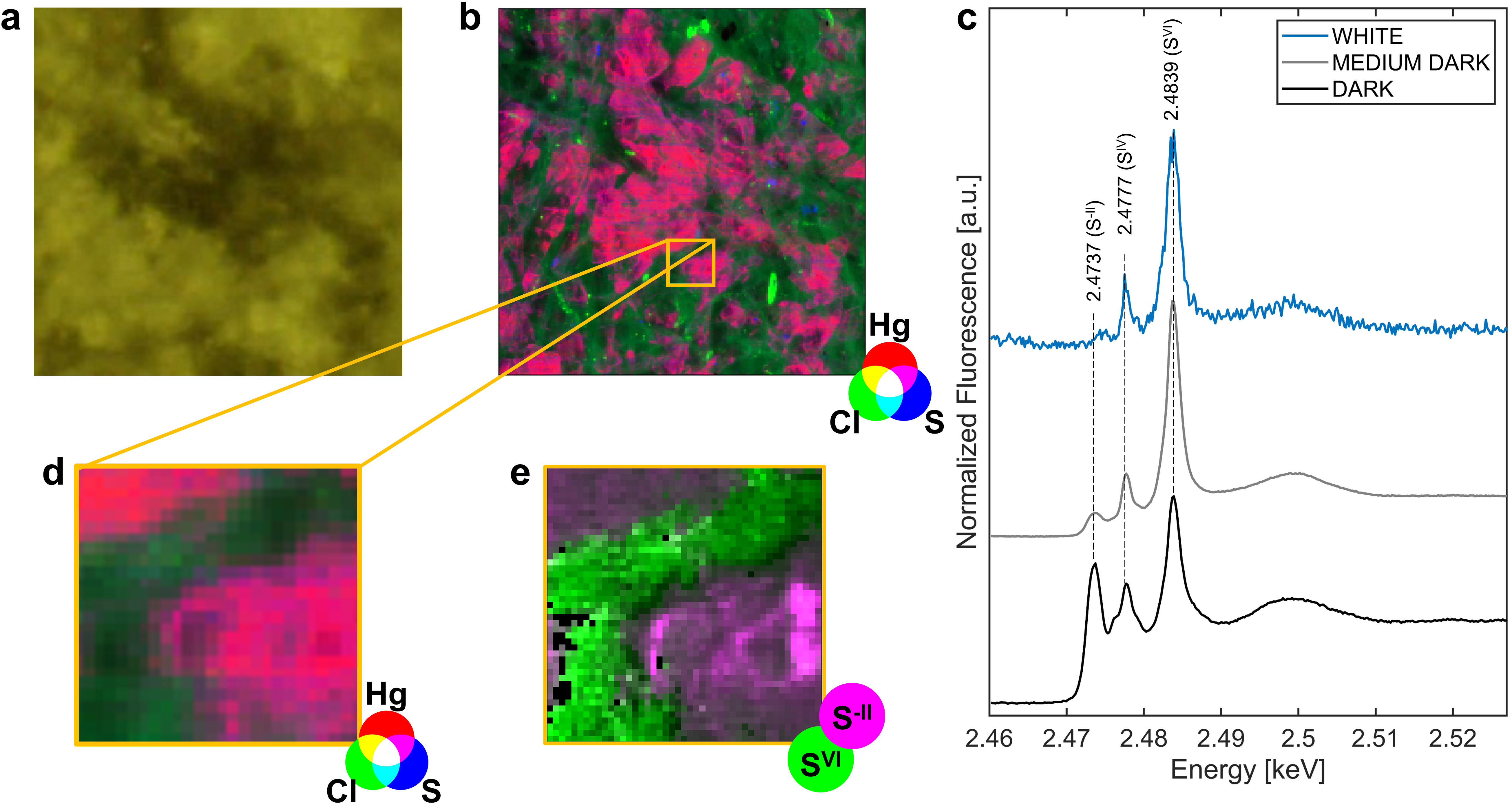
A variety of synchrotron radiation-based X-ray investigations were carried out to characterise the small particles that made up the dark stains. Synchrotron-based X-ray fluorescence (XRF) mapping and X-ray absorption near-edge structure (XANES) data were acquired at beamline ID21. Macro and µXRF maps supported earlier findings from X-ray spectroscopy analyses, inferring the presence of Hg, S, Ca, Al, Cl, Si and K. Mercury was detected in the darkened region, while it was not detected in the region without alteration. A similar distribution was found for sulfur, although it was also detected in the white regions. µXRF mapping of a stain showed a co-localisation of Hg and S that follows the shape of the dark stain almost perfectly (Figure 1a,b,d).
Click image to enlarge
Fig. 1: a) Visible light image of the black stain region region where µXRF mapping was performed [step size (h × v), 4 × 4 μm; map size (h × v), 800 × 800 μm; exp. time, 100 ms/pixel; energy: 2.825 keV]. b) RGB composite of µXRF maps of Hg (red), Cl (green) and S (blue). The magenta colour indicates the region where mercury and sulfur signals overlap, following the shape of the black stain. c) Macro-XANES spectra recorded at the S K-edge. Peak at 2.4737 keV corresponds to the sulfides (S-II), peak at 2.4777 keV to the sulfites (SIV) and peak at 2.4839 keV to the sulfates (SVI). d) Detail of RGB composite of µXRF maps of Hg (red), Cl (green) and S (blue) [step size (h × v), 4 × 4 μm]. e) Selected area of interest for hyperspectral 2D µXRF maps at S K-edge. The RGB composite image shows the distribution of sulfides (S-II) and sulfates (SVI) species.
Macro-XANES spectra acquired at the S K-edge (Figure 1c) exhibited a remarkable difference between darkened and non-altered areas, both in signal intensity (high in dark, low in white region) and sulfur oxidation states (mainly sulfates (SVI) and sulfites (SIV) in white, also sulfides (S-II) in the dark region), suggesting a predominant formation of sulfides associated with the blackening (Figure 1e).
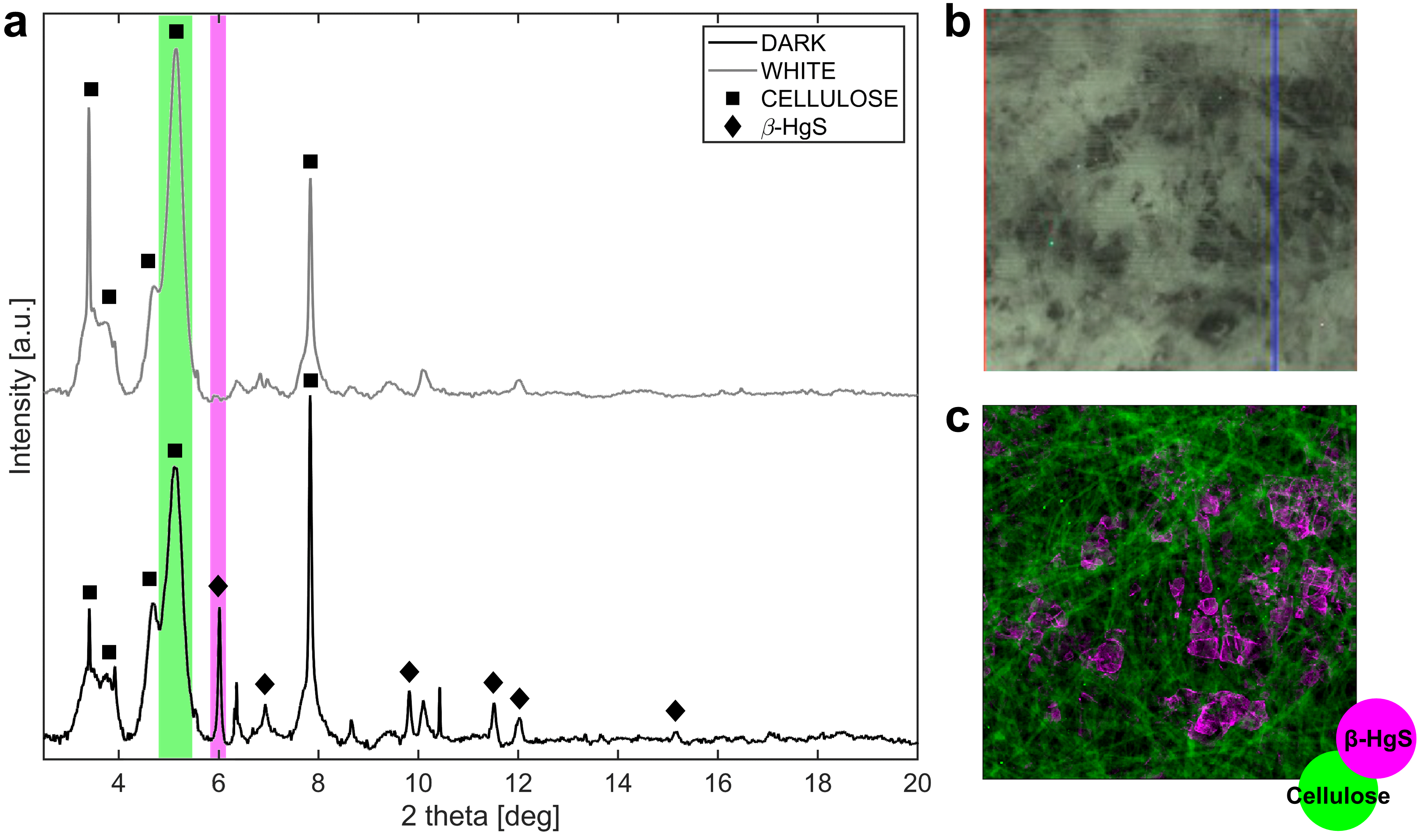
Two types of X-ray diffraction (XRD) measurements were performed within the framework of the ESRF’s “Historical Materials block allocation group (BAG)” beamtime access (www.esrf.fr/BAG/HG172). High angular resolution XRD (HR-XRD) measurements were collected at beamline ID22 on samples from the dark and white regions, and μXRD maps were acquired at beamline ID13. Thanks to these observations, the spatial distribution of the black stains and metacinnabar (β-HgS) peak heights could be correlated (Figure 2).
Click to enlarge
Fig. 2: a) HR-XRD spectra patterns of the white (grey) and dark (black) regions. Squares and diamonds highlight the peaks related to cellulose and β-HgS respectively. b) Visible light photograph of the area of the dark sample where µXRD mapping was performed [step size (h × v), 3 × 3 μm; map size (h × v), 1.2 × 1.2 mm; exp. time, 10 ms/pixel; energy: 13 keV]. c) µXRD images calculated by integrating XRD intensity over regions of interest of XRD pattern, shown as green and purple rectangles in (a) for cellulose and metacinnabar respectively.
The research highlighted evidence of the spontaneous formation of β-HgS particles in conservation conditions (i.e., controlled environmental conditions including absence of light and low humidity). A first hypothesis about Hg provenance is based on the possible use of anti-vegetative mercury salts (HgCl2) in the gluing mixture. However, it is important to stress that this compound was not found throughout the current research. Additionally, it remains to determine if paper hydrolysis and the degrading processes of the glue mixture revealed by ATR-FTIR imaging may have contributed to the blackening phenomenon. Further studies are required to propose a robust mechanism of the formation of β-HgS and to verify if the exposure of HgCl2 to different sulfur sources, in mild environmental conditions, could lead to the formation of metacinnabar black particles.
Principal publication and authors
Imaging and micro-invasive analyses of black stains on the passepartout of Codex Atlanticus Folio 843 by Leonardo da Vinci, N. Guarnieri (a), M. Ghirardello (b), S. Goidanich (a), D. Comelli (b), D. Dellasega (c), M. Cotte (d,e), E. Fontana (f), L. Toniolo (a), Sci. Rep. 13, 4902 (2023); https://doi.org/10.1038/s41598-023-31129-2
(a) Department of Chemistry, Materials and Chemical Engineering, Politecnico di Milano, Milan (Italy)
(b) Department of Physics, Politecnico di Milano, Milan (Italy)
(c) Department of Energy, Politecnico di Milano, Milan (Italy)
(d) ESRF
(e) Laboratoire d’Archéologie Moléculaire et Structural (LAMS) CNRS UMR 8220, Sorbonne Université, Paris (France)
(f) Veneranda Biblioteca Ambrosiana, Milan (Italy)
| About the beamlines |
| ID13: ID13 is dedicated to high-spatial-resolution diffraction and scattering experiments using focused monochromatic X-ray beams. Two endstations, a microbranch and a nanobranch, are operated in time-sharing mode. Scanning diffraction and scattering experiments are performed in both branches, often combined with simultaneous scanning X-ray microfluorescence. A broad range of materials can be examined, from bio-materials, biological tissues and synthetic polymers to inorganic materials, e.g., for sustainable energy applications. The current set-ups allow to examine single crystals (including proteins), fibres or other extended samples. The availability of microbeams has led to the development of specific sample environments and geometries including stretching and nano-indentation cells, humidity control, chip-based nanocalorimetry, micro-grazing-incidence scattering/diffraction (GISAXS/GIWAXS), and microfluidics environments. |
|
ID21: ID21 is an X-ray micro-spectroscopy beamline dedicated to micro X-ray fluorescence (μXRF) and micro X-ray absorption near-edge structure (μXANES). The scanning X-ray microscope can be operated in an energy range from 2-11 keV, thus giving access to the K-edges of phosphorus to zinc, and to the L- and M-edges of some heavier elements. The ID21 microscope is regularly used to study ancient and artistic materials. Analyses can be carried out on almost all kinds of artistic materials, such as paintings, ceramics, glasses, plastics, wood and papyrus, shedding light on degradation mechanisms and ultimately helping to optimise strategies for the preservation and conservation of cultural heritage objects. ID21 has also been a pioneer in the use of X-ray microspectroscopy applied to environmental and earth sciences. |
| ID22: ID22 is a high-resolution powder diffraction beamline that allows the identification of different crystalline materials even if present in a sample at low concentration. Each crystalline substance has its own distinct diffraction pattern, like a fingerprint, and the high quality of the data from ID22 makes it possible to identify what is present even from minute amounts of a particular substance, or to disentangle the individual components of a complex mixture of many substances. Materials that can be studied in this way range from substances that are associated with the fabrication, degradation and conservation of art treasures, to contaminants in pharmaceutical active ingredients, or trace phases influencing mechanical properties in metallurgical samples such as steel. |
partners


European Synchrotron Radiation Facility - 71, avenue des Martyrs, CS 40220, 38043 Grenoble Cedex 9, France.