- Home
- News
- Spotlight on Science
- Multiscale X-ray...
Multiscale X-ray analysis reveals mineral phase shifts in bone cancer subtypes
25-07-2025
X-ray absorption near-edge structure spectroscopy and X-ray fluorescence imaging at beamline ID21 reveal disruptions in mineral structure across osteosarcoma subtypes. Combined with X-ray diffraction and microcomputed tomography, these approaches elucidate tumour–bone interactions and open new avenues for diagnostics and therapeutic monitoring.
Share
Osteosarcoma (OS) is the most common primary bone cancer in children and adolescents, and poses a significant challenge in paediatric oncology. Despite decades of research, survival rates remain unchanged, largely due to the tumour’s extensive heterogeneity and limited insights into how it disrupts the surrounding tumour microenvironment [1]. While OS is known to alter bone remodelling and mineralization, few studies have explored these effects at the molecular and structural levels in human tissue [2,3].
In this study, researchers investigated the impact of three OS subtypes on the morphology, architectural structure, and mineral composition of human bone: high-grade osteoblastic OS (HG-OS), the most common and aggressive subtype, which produces malignant immature tissue within the bone; periosteal OS (PA-OS), a slow-growing subtype forming dense ossified masses on the surface of the periosteum; and periosteal OS (PE-OS), an intermediately aggressiveness subtype in which cartilage and bone develop on the inner periosteal layer. The study focused on how tumour-driven remodelling alters bone-tissue architecture and hydroxyapatite (HA), the primary bone mineral, while aiming to detect compositional changes undetectable by conventional histology, using adjacent non-tumour tissues as internal controls.
To achieve this goal, multiscale imaging was performed on paired tumour and control bone samples from the same patients. X-ray computed microtomography (XCMT) at Elettra synchrotron in Italy was combined with micro X-ray absorption near-edge spectroscopy (µXANES) at the Ca K-edge and micro X-ray fluorescence microscopy (µXRF), both at ESRF beamline ID21, to probe the chemical state of calcium in the bone mineral phase. Additionally, X-ray diffraction (XRD) at Elettra assessed changes in the HA crystal structure. Resected bone tissues from tumour and adjacent control regions were preserved at –20°C. XCMT scans were performed on the frozen samples, which were then dehydrated, infiltrated with resin, and sectioned into 60 - 100 µm slices for µXANES and µXRD analyses.
The results demonstrate that OS impacts bone mineralization in a subtype-dependent and structurally complex manner. Morphologically, PA-OS and PE-OS exhibited distinct alterations compared with each other and with control tissues, whereas HG-OS resemble normal bone. XRD revealed c-axis distortions in the PA-OS and PE-OS, indicative of crystal strain in their HA phases.
Click figure to enlarge
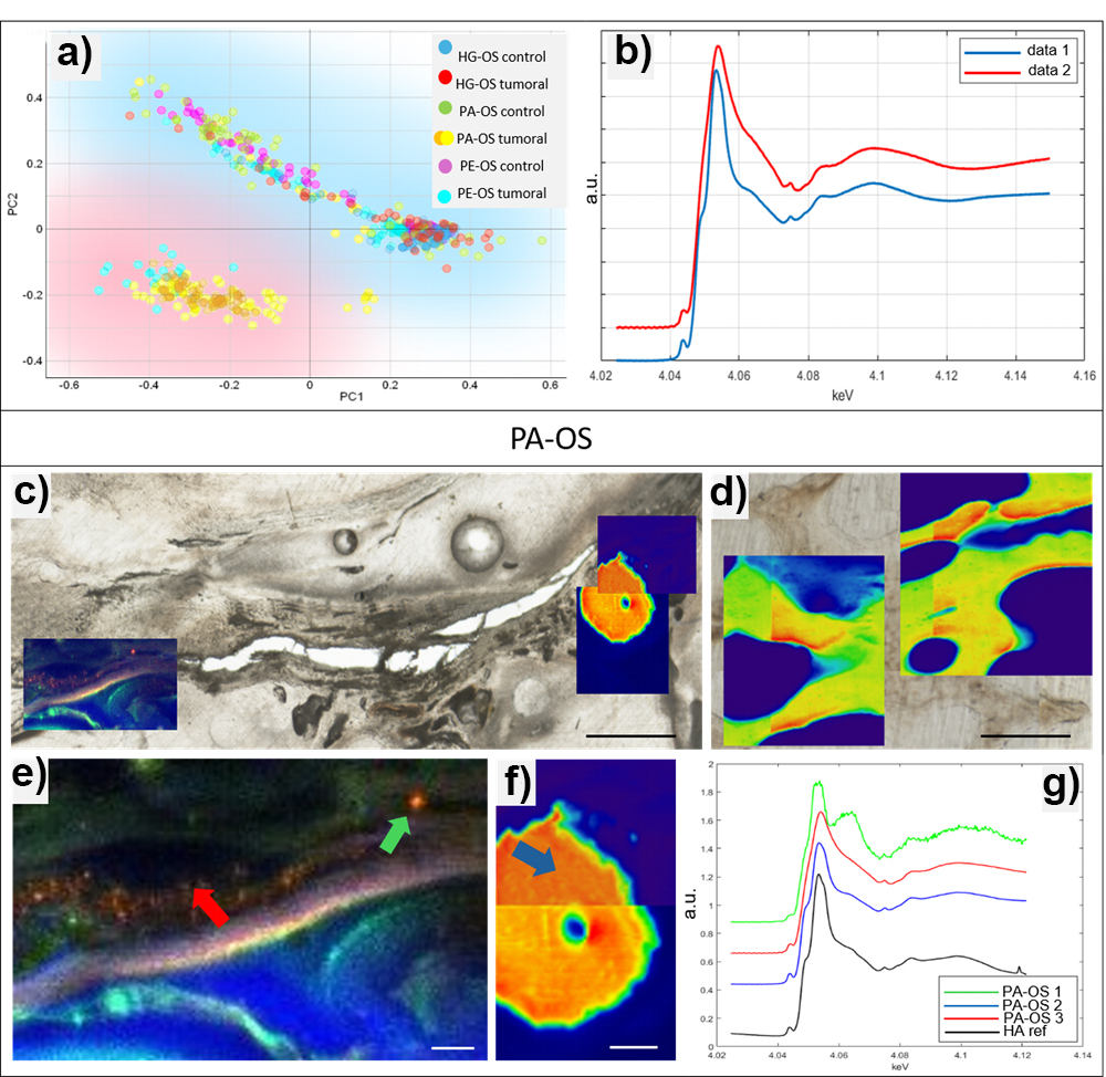
Fig. 1: Principal component analysis (PCA) results. a) Scatter plot of PC1 versus PC2 showing two distinct clusters (blue and red). b) Mean µXANES spectra from the blue cluster (“data 1”) and the red cluster (“data 2”). c,d) XRF elemental maps of a PA-OS sample and a control sample, respectively (scale bar: 1 mm). e,f) Magnified views of the ROI in (c): in (e), elemental overlays (Ca = red, P = blue, K = green); in (f), Ca map only (scale bar: 0.2 mm). g) µXANES spectra from the points indicated by arrows in (e) and (f): PA-OS 1 (green), corresponding to amorphous calcium carbonate; PA-OS 2 (blue), from structured bone tissue; PA-OS 3 (red), from disorganized bone tissue. The hydroxyapatite reference spectrum is shown in black.
To further dissect subtype effects on mineral chemistry, µXRF mapped key elements (P, K, Ca) at the micrometre scale, and µXANES probed calcium speciation in selected regions. Principal component analysis (PCA) of µXANES spectra yielded two clusters (Figure 1a): control and HG-OS (blue) versus PE-OS and PA-OS (red). HG-OS spectra matched the HA reference (data 1 in Figure 1b), signifying mature mineral, while PE-OS and PA-OS spectra deviated from HA, suggesting amorphous calcium carbonate (ACC) presence (data 2 in Figure 1b).
XRF enabled detection of both HA-like and ACC-like mineral domains. ACC-like signatures appeared in disorganized tumour regions (Figure 1c, red arrow in Figure 1e and red spectrum in Figure 1g), whereas adjacent control bone exhibited spectra matching HA (Figure 1d). Within tumour tissue, XRF mapping revealed distinct P- and K-rich areas, reflecting an organized organic matrix, coexisting with calcium carbonate signals (Figure 1e and green spectrum in Figure 1g). Conversely, spectra from more structurally intact tumour zones (Figure 1f and blue spectrum in Figure 1g) overlapped closely with the HA reference (Figure 1g, black spectrum). These observations define a subtype-specific biomineralization pattern in OS: HG-OS maintains a mature HA signature, while PA-OS and PE-OS show heterogeneous mineralization, combining crystalline HA and amorphous calcium phases.
This study underscores the power of a synchrotron-based, multiscale imaging approach to reveal nuanced features of the tumour microenvironment. By comparing paired tumour and control samples from the same patient, interindividual variability and isolate disease-driven changes can be effectively controlled. Although constrained by OS rarity and sample availability, the current findings pave the way for larger-cohort studies. Revealing subtle shifts in mineral composition and crystal lattice provides a foundation for developing mineral-based biomarkers, guiding therapeutic monitoring, and ultimately improving patient outcomes in this aggressive cancer.
Principal publication
Insights into the osteosarcoma microenvironment: Multiscale analysis of structural and mineral heterogeneity, F. Rossi et al., Acta Biomaterialia 199, 193-201 (2025); https://doi.org/10.1016/j.actbio.2025.04.057
References
[1] I. Corre et al., Cells 9, 976 (2020).
[2] B. Zanghellini et al., J. Struct. Biol. 216, 18106 (2024).
[3] B. Zanghellini et al., J. Struct. Biol. 207, 55 (2019).
| About the beamline: ID21 |
|
The ID21 beamline is dedicated to micro and nano-X ray spectroscopy, offering 2D X-ray fluorescence (XRF) mapping and X-ray absorption spectroscopy (XAS) in the tender X-ray energy range (2.1 - 10.5 keV). These techniques can be combined to produce multi-energy XRF maps, enabling both 2D elemental mapping and chemical speciation in 0D, 1D, and 2D. The beamline is optimized for the detection and chemical analysis of elements from phosphorus to zinc, while heavier elements can also be studied via their L- and M-edge absorption.
|
partners


European Synchrotron Radiation Facility - 71, avenue des Martyrs, CS 40220, 38043 Grenoble Cedex 9, France.