- Home
- News
- General News
- Trilobite’s last...
Trilobite’s last meal revealed by synchrotron microtomography
11-10-2023
The gut contents of a 465 million-year-old fossilised trilobite were imaged at the ESRF using synchrotron microtomography technique. The results, published in Nature, shed light on the feeding habits and lifestyle of one of the most common and well-known fossil arthropods. The research fills a fundamental gap in the understanding of trilobite ecology and their role in Paleozoic ecosystems.
Share
Trilobites are among the most iconic of fossils and formed a highly diverse, abundant and important component of marine ecosystems during most of their 270-million-year-long history from the early Cambrian period to the end Permian period. More than 20,000 species have been described to date.
Despite numerous fossil specimens, the feeding habits of these animals have had to be inferred indirectly, because no known fossil specimens with internal gut contents have previously been reported. This knowledge gap limits the ability to understand trilobites’ ecological roles, which in turn affects the overall understanding of the ecosystems that they inhabited. A specimen of the trilobite Bohemolichas incola, with partly visible shelly gut contents, was present in a Czech public collection, but the inability to image and identify the individual shell fragments without destroying the fossil limited its research potential.
A team of researchers led by Per Erik Ahlberg at Uppsala University, Sweden, and Valéria Vaškaninová at Charles University, Czech Republic, came to the ESRF to investigate this rare fossil using propagation phase-contrast synchrotron microtomography, at ESRF ID19 beamline. The technique enabled the scientists to non-destructively image all the shell fragments in the gut in 3D and at high resolution. The result was a complete map showing the position and identity of each shell fragment in the gut.
"ESRF played an absolutely pivotal role in this study" says lead author Per Ahlberg, professor at Uppsala University. "The resolution and scan quality it provides - this scan was made on the old ID19 beamline before the upgrade, even better results are possible now - were essential to identify the gut contents, piece by piece. A conventional CT scan would have told us that the trilobite had been eating; only ESRF could tell us what it had been eating."
 |
 |
|
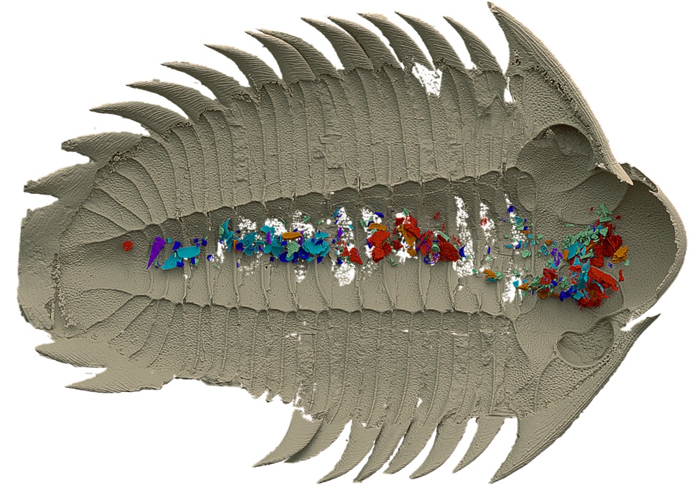
Scanned model of the specimen in ventral view, with the exoskeleton modelled in tan and the hypostome (a hard mouthpart of trilobites) in yellow (left image). The digestive-tract contents are separated and colour-coded into various fossil groups. Scale bar, 10 mm.Credit: Kraft, P. et al./Nature (CC BY 4.0) |
|
The study demonstrates that the trilobite had been eating a range of small benthic invertebrates (which live on the sea bed), including ostracods (small crustaceans with paired shells), echinoderms (animals related to modern starfish and sea urchins) and hyolithids (cone-shaped animals with no living representatives). The results indicate also that its gut had a neutral to alkaline pH. The authors propose that Bohemolichas incola was an opportunistic scavenger. It was a light crusher and a chance feeder that ate dead or living animals, which either disintegrated easily or were small enough to be swallowed whole. After death, this scavenger became scavenged. The fossil specimen also shows the vertical tracks of other scavengers that burrowed into the trilobite’s carcass, where they targeted the soft tissue but avoided the gut. This implies noxious conditions inside the trilobite’s digestive system and possible ongoing enzymatic activity.
The findings demonstrate key aspects of trilobite biology that have previously been suggested, but never illustrated, and it fills a fundamental gap in the understanding of the role of trilobites in Paleozoic ecosystems.
Reference:
Top image: The trilobite Bohemolichas incola. Credits: Jiri Svoboda
partners


European Synchrotron Radiation Facility - 71, avenue des Martyrs, CS 40220, 38043 Grenoble Cedex 9, France.