- Home
- News
- General News
- Breast cancer scans...
Breast cancer scans possible with a 25 times reduced radiation dose
19-10-2012
Synchrotron X-rays confirm a new method for X-ray image processing.
Share
Joint release by the ESRF and LMU and UCLA.
Grenoble, 22 October 2012: Scientists have developed a way to produce three-dimensional X-ray images of the breast at a radiation dose that is lower than the 2D radiography methods used in clinics today. The new method enables the production of 3D diagnostic computed tomography (CT) images with a spatial resolution 2-3 times higher than present hospital scanners, but with a radiation dose that is about 25 times lower. This breakthrough has the potential to overcome the main obstacle limiting conventional CT imaging of the breast: the high radiosensitivity of the breast glandular tissue. Synchrotron X-rays at beamline ID17, the medical station of the ESRF, have been used to test the technique which, once deployed in hospitals, will make CT scans a diagnostic tool to complement dual view mammography.
The results of this innovative method are published in the online Early Edition of the Proceedings of the National Academy of Sciences (PNAS) for the week beginning 22 October 2012.
The multidisciplinary team comprised physicists, radiologists and mathematicians from the European Synchrotron Radiation Facility (ESRF, Grenoble, France), the Ludwig Maximilians University in Munich (LMU, Cluster of Excellence MAP) and the University of California at Los Angeles (UCLA). The first authors are Yunzhe Zhao of UCLA and Emmanuel Brun of the LMU/ESRF.
Early detection contributes to an improved prognosis and results in reduced breast cancer mortality. The breast cancer screening method typically used today is “dual-view digital mammography”. The limitation is that it only provides two images of the breast tissue, which can explain why 10 percent to 20 percent of breast tumours are not detectable on mammograms. Mammograms can also sometimes appear abnormal, when no breast cancers are actually present.
Computed tomography (CT), an X-ray technique that allows a precise 3D visualisation of the human body organs, cannot be routinely used for breast cancer diagnosis because the risk of long-term effects in radiosensitive organs like the breast is considered too high.
Recognising these limitations, scientists have tackled the problem using a new approach. They combined three ingredients that together should now make CT scans for early detection of breast cancer become possible. These ingredients are: high energy X-rays, a special detection method called “phase contrast imaging” and the use of a sophisticated novel mathematical algorithm, known as “equally sloped tomography” (EST), to reconstruct the CT images from X-ray data. Tissues are more transparent to high energy X-rays and therefore less dose is deposited (a factor of 6 in radiation dose reduction). Phase contrast imaging, mastered by the ESRF and the LMU-MAP teams, allows the production of images using fewer X-rays to obtain the same image contrast. Finally, the EST method, originally developed by researchers at UCLA, needs 4 times less radiation to obtain the same image quality.
The team X-rayed a human breast at multiple different angles using phase contrast tomography and applied the EST algorithm to 512 images to produce higher resolution 3-D images of the organ than ever before and at a lower dose than a mammogram.
 |
|
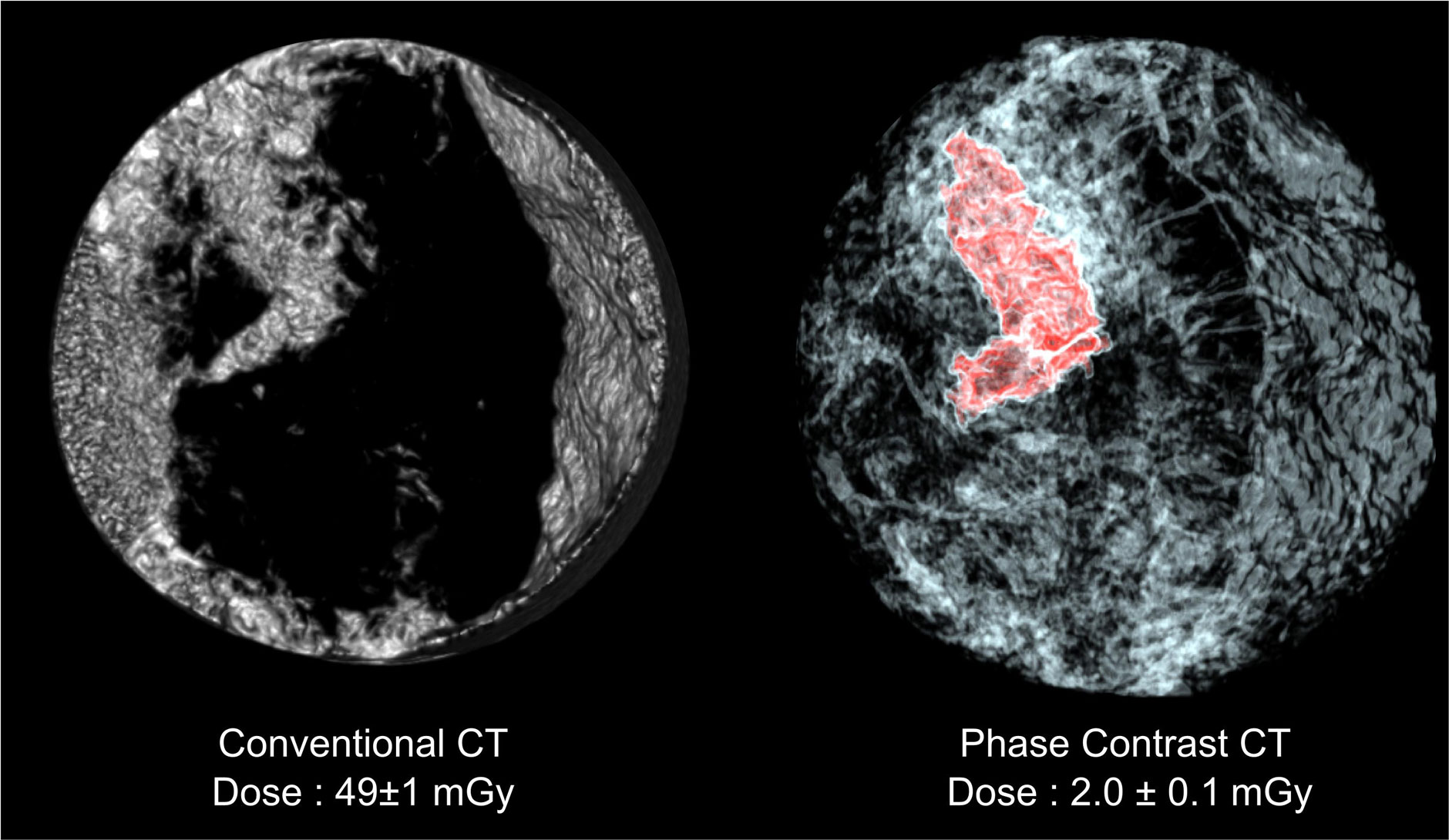
Comparison between a conventional CT scan of the breast sample and a scan using equally sloped tomography with phase contrast imaging. In the latter image, the tumour is highlighted in red. The radiation dose needed for the scans is indicated at the bottom. Credit ESRF-LMU/Brun. |
In a blind evaluation, five independent radiologists from the LMU ranked the generated images as having the highest sharpness, contrast, and overall image quality compared to 3-D images of breast tissue created through other standard methods.
“This new technique can open up the doors to the clinical use of computed tomography in the breast diagnosis, which would be a powerful tool to fight even better and earlier against breast cancer”, says Prof. Maximilian Reiser, Director of the Radiology Department of the LMU, which provided the medical expertise for this research. “This result has been obtained thanks to the synergy of the expertise by researchers from very different disciplines. These high-quality X-ray CT images at high energies are the result of a 10-year effort at the ESRF” says Alberto Bravin, head of the ESRF medical research laboratory who led the team in Grenoble. “After dramatically reducing the dose delivered during the examination of the breast, our next objective is to develop this technique in the early visualisation of other human diseases and to work towards its clinical implementation.” adds Paola Coan, Professor of X-ray imaging at the LMU and member of the Munich-Centre for Advanced Photonics (MAP), who led the group from Munich.
 |
|
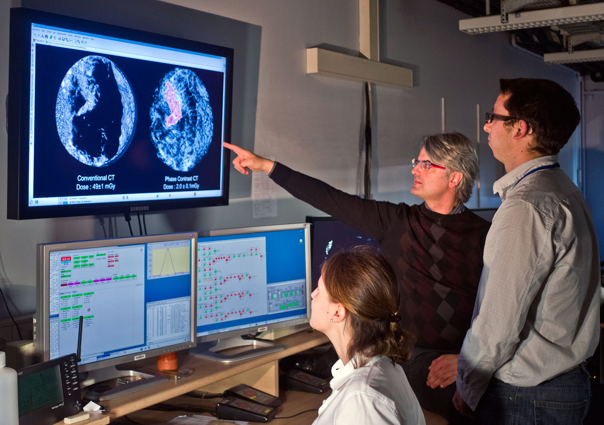
Photo of the Grenoble team in the control room of the medical research laboratory. From left to right: Paola Coan, Alberto Bravin and Emmanuel Brun. Credit ESRF/Blascha Faust. |
“Three-dimensional reconstructions, like the ones created in this research, are produced using sophisticated software and a powerful computer that can combine many images into one 3-D image, much like the slices of an orange. By rethinking the mathematic equations of the software in use today” says Jianwei (John) Miao, UCLA professor of physics and astronomy and researcher with the California NanoSystems Institute at UCLA “we developed a more powerful algorithm that requires fewer slices to get a clearer 3-D picture”.
Today, the new technology is in the research phase and will not be available to patients for some time. To be implemented in clinics, it needs an X-ray source small enough to become commonly used for breast cancer screening. “Many research groups are actively working to develop this device and once this hurdle is cleared, the new X-ray technique is poised to make a big impact on society”, concludes Emmanuel Brun.
A video of the CT scan is available on YouTube: 3-D CT scan of breast sample using equally sloped tomography with phase contrast imaging.
Reference
Y. Zhao, E. Brun et al., "High resolution, low dose phase contrast X-ray tomography for 3D diagnosis of human breast cancers", PNAS, early online edition, week starting 22 October 2012.
Top image: Phase contrast CT scan of a breast sample. Credit ESRF-LMU/Brun.
partners


European Synchrotron Radiation Facility - 71, avenue des Martyrs, CS 40220, 38043 Grenoble Cedex 9, France.