Enhanced crystal characterization with Rocking Curve Imaging at BM05
Emerging X-ray free-electron laser (XFEL) techniques, such as X-ray multiple projection imaging, necessitate exceptionally high-quality crystals. To meet these requirements, the Rocking Curve Imaging (RCI) technique implemented at the BM05 beamline has demonstrated its ability to provide precise crystal characterization, offering the required field of view, spatial resolution, and angular resolution.
Share
X-ray multiple projection imaging (XMPI) is an innovative technique that capitalizes on the advanced capabilities of modern synchrotron and XFEL sources. This method employs a beam-splitting scheme to achieve simultaneous illumination of a sample from multiple directions, enabling the study of dynamic processes without sample rotation. However, the successful implementation of XMPI depends on the perfection of the crystals used in its optical setups. Consequently, high-resolution crystal characterization is essential prior to their application.
The ESRF has developed a quantitative Bragg diffraction imaging method, referred to as Rocking Curve Imaging (RCI), at the BM05 beamline. This technique, often termed ‘X-ray topography’, enables the characterization of crystal properties over several square millimetres. With micrometre spatial resolution and microradian angular resolution, RCI provides detailed maps of the “local” diffracted integrated intensity, peak width (full width at half maximum, FWHM), and peak position. These high-resolution maps facilitate the detection and quantification of crystalline imperfections, such as dislocations, surface defects, and distortions.
RCI was employed to assess the suitability of diamond, silicon, and germanium single crystals intended for use as “splitters” or “recombiners” in XMPI optical setups. These crystals are designed to prevent clamping-related distortions from propagating into the diffraction region, which must remain free of surface or crystalline defects. High-resolution RCI maps (1.3 x 1.3 mm2, pixel size 0.65 µm) of these crystals clearly detected and quantified residual distortion, scratches and crystalline defects.
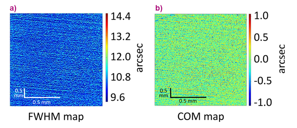
The diamond splitters were shown to meet the required quality standards, with a small number of defects within acceptable tolerances. They exhibited a local FWHM of approximately 20 µradian and a peak position variation of less than 2 µradian across the entire investigated area. In contrast, the germanium recombiners, while displaying uniform and relatively good overall quality, were found to have a noticeably distorted surface, with FWHM values in the range of 40-70 µradian and a peak position variation of approximately 5 µradian, as illustrated in Figure 1. These distortions may be attributed to the brittle structure and lower hardness of germanium, as well as to the comparatively less advanced finishing technologies available for this material relative to silicon or diamond.
Click figure to enlarge
Fig. 1: Monochromatic high-resolution X-ray topography of germanium recombiners in parallel geometry, performed at the BM05 beamline. The analysis was based on Bragg diffraction from the (440) lattice planes of the recombiners. The images cover a crystal surface area of 1.3 mm wide (horizontal) and 5 mm long (vertical), the latter corresponding to the elongated beam footprint in the diffraction direction. The photon energy was 20 keV, with a field of view of 1.3 mm × 1.3 mm and a pixel size of 0.65 µm. a) Full width at half maximum (FWHM) map of the surface, representing the diffraction passband at each point. b) Centre-of-mass (COM) map, indicating the relative position of the centre of the rocking curve at each surface point.
Most investigations conducted at BM05 using RCI focus on crystals intended for applied purposes, where this quantitative Bragg imaging method provides a unique and effective characterization tool. More broadly, Bragg diffraction imaging enables the detailed analysis of defects in high-quality semiconductor crystals and crystalline layers, with micrometre-scale spatial resolution and microradian-scale angular resolution. RCI thus offers significant value for the characterization and optimization of crystals and deposited layers.
Principal publication and authors
Development of crystal optics for X-ray multi-projection imaging for synchrotron and XFEL sources, V. Bellucci (a), S. Birnsteinova (a), T. Sato (a), R. Letrun (a), J.C.P. Koliyadu (a), C. Kim (a), G. Giovanetti (a), C. Deiter (a), L. Samoylova (a), I. Petrov (a), L. Lopez Morillo (a), R. Graceffa (a), L. Adriano (a), H. Huelsen (b), H. Kollmann (b), T.N. Tran Calliste (c), D. Korytar (d), Z. Zaprazny (e), A. Mazzolari (f,g), M. Romagnoni (f,g), E. Myrto Asimakopoulou (h), Z. Yao (h), Y. Zhang (h), J. Ulicny (i), A. Meents (j), H.N. Chapman (j,m), R. Bean (a), A. Mancuso (a,k,l), P. Villanueva-Perez (h), P. Vagovica (j), J. Synchrotron Radiat. 31, 1534-1550 (2024); https://doi.org/10.1107/S1600577524008488
(a) European XFEL GmbH, Schenefeld (Germany)
(b) SmarAct GmbH, Oldenburg (Germany)
(c) ESRF
(d) Integra TDS Ltd, Piestany (Slovakia)
(e) Institute of Electrical Engineering, Bratislava (Slovakia)
(f) University of Ferrara, Ferrara (Italy)
(g) INFN – Istituto Nazionale di Fisica Nucleare, Ferrara (Italy)
(h) Synchrotron Radiation Research and NanoLund, Lund University (Sweden)
(i) University of P. J. Safarik, Kosice (Slovakia)
(j) Center for Free-Electron Laser Science (CFEL), DESY, Hamburg (Germany)
(k) Diamond Light Source, Harwell Science and Innovation Campus, Didcot (UK)
(l) Department of Chemistry and Physics, La Trobe Institute for Molecular Science, La Trobe University, Melbourne, Victoria (Australia)
(m) University of Hamburg, Hamburg (Germany)
partners


European Synchrotron Radiation Facility - 71, avenue des Martyrs, CS 40220, 38043 Grenoble Cedex 9, France.