- Home
- News
- Spotlight on Science
- Self-transformation...
Self-transformation of solid CaCO3 microspheres into core-shell and hollow hierarchical structures
27-10-2022
Coherent X-ray diffraction and fluorescence imaging techniques at beamlines ID10 and ID16 were used to reveal the self-transformation mechanisms of solid vaterite microparticles stabilised by polystyrene sulfonate into macroporous, core-shell or hollow microspheroids.
Share
The common denominator between ice cream, solar cells and an aluminium alloy used in the fuselage of aircraft is that all these multiphase systems evolve during prolonged ageing according to a mechanism called Ostwald ripening, involving the transport of matter from smaller crystals to larger ones. Ostwald ripening under controlled manipulation can be fruitfully used to obtain hollow microspheres from solid ones. However, the formation of such complex structures cannot be explained by Ostwald ripening alone.
The self-transformation of solid CaCO3 microspheres [1] into complex core-shell and hollow particles was therefore investigated by coherent X-ray diffraction (CXDI) [2] and 3D X-ray fluorescence imaging (3D-XRF) [3] at beamlines ID10 and ID16A. The techniques were used to visualise in 3D the time-dependent formation of hollow microparticles of calcium carbonate in the presence of polystyrene sulfonate (PSS).
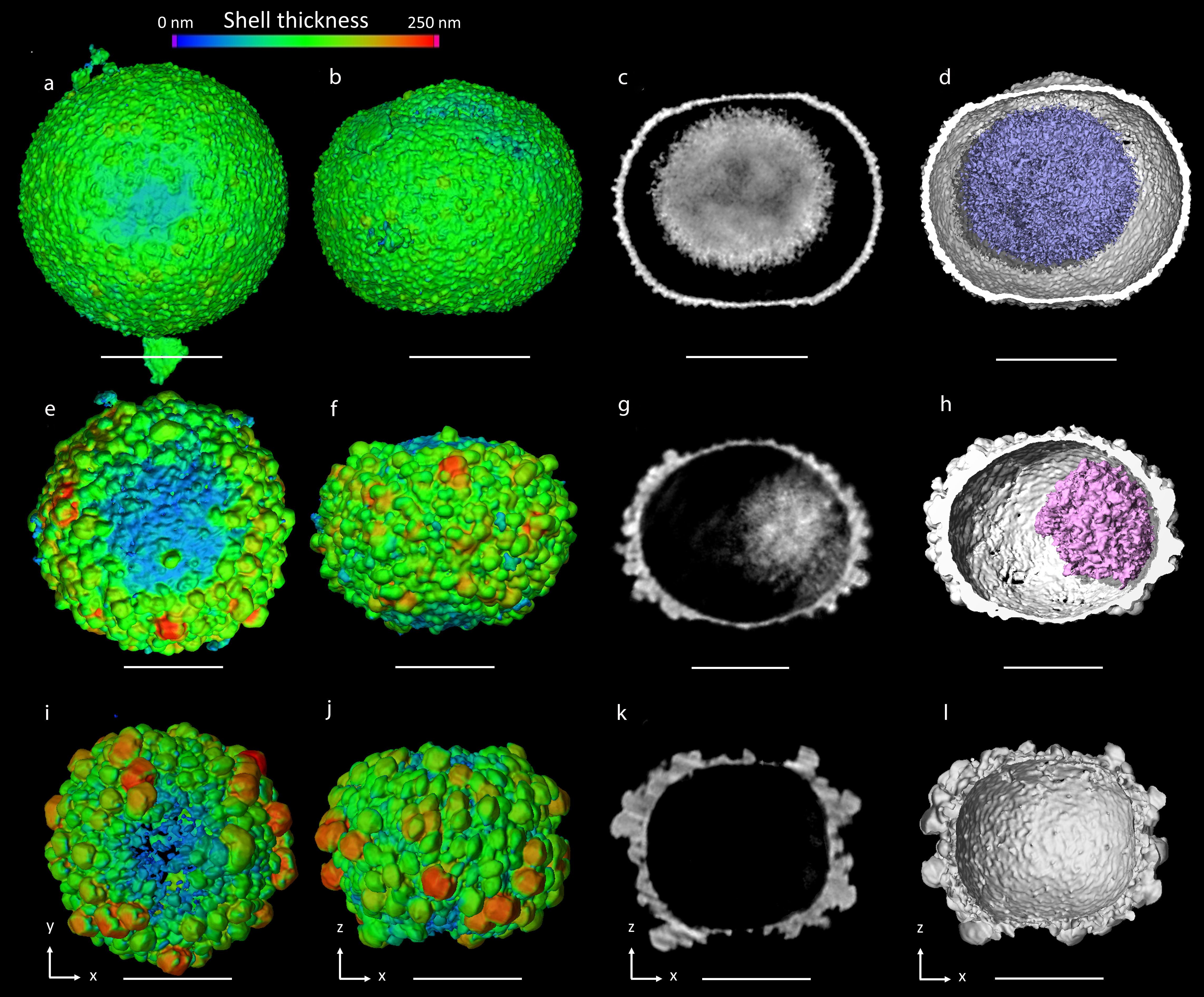
The results suggest that the formation of a gap between the shell and the core takes place because the nanoparticles in the inner part of the shell are more stable than those of the core, because of their higher sizes or the higher content of PSS, or both (Figures 1a-d). After 8 hours of reaction, the core becomes drastically smaller due to progressive dissolution with time, concomitantly with the growth of vaterite crystals on the outer surface of the shell (Figures 1e-h). After 24 hours of reaction, the final hierarchical structure of the microparticle is characterised by a completely hollow core, a shell with an oblate spheroidal morphology, and with thicknesses between 0 in the region of the poles and around 250 nm at the equator (Figures 1i-l). The outer surface of the shell is decorated by crystals of a few hundred nanometres in size, in the form of hexagonal-based bipyramids, mainly oriented along the meridians.
Click image to enlarge
Fig. 1: CXDI images of vaterite oblate microspheroids obtained from the syntheses of 8mM of CaCl2, 8mM of Na2CO3 and 1g/L of PSS after (a-d) 2h of reaction, (e-h) 8h of reaction and (i-l) 24h of reaction. a), (b), (e), (f), (i) and (j) 3D views showing the wall thickness of the shell from 0 to 250 nm. c), (g) and (k) CXDI 2D slices viewed in the xz plane. d) and (h) 3D views of the core depicted in blue, with half the shell cut away to allow observation of the inside. Scale bar = 2 µm for (a-d) and 1 µm for (e-l).
Varying the initial concentration of the precursors (Ca2+, carbonate, and/or PSS) demonstrated that a hollow core is not necessarily obtained. When the initial concentration of PSS is high, it inhibits the dissolution of the nanocrystals of vaterite but also their growth by Ostwald ripening.
Click image to enlarge
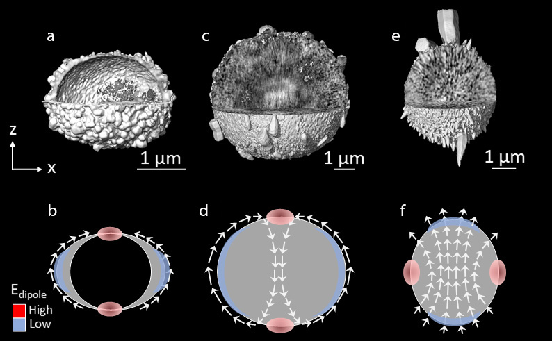
Fig. 2: Nanoparticle dipole order controls the shape of the microspheroids. Microspheroids (a,b) 8mM-1gL-8h, (c,d) 30mM-0.24gL-24h and (e,f) 120mM-1gL-8h. Images (a), (c) and (e) obtained by CXDI. b,d,f) Drawings representing 2D views of the microspheroids shown in (a), (c) and (e) respectively. White arrows illustrate the orientation of c-axis of the nanocrystals. The red ellipse and blue lunules refer to regions of high and low dipole energy respectively.
In addition, the data indicate a link between the flattened morphology of the shell, the presence of holes at the surface of the shell, and the co-alignment of the nanocrystals composing the shell (Figure 2). The core/shell microspheroids shown in Figures 1 and 2a are oblate because the c-axes of the nanocrystals are in the plane of the shell. This is represented in Figure 2b, where white arrows display the orientation of the c-axis. It is proposed that the in-plane alignment of the nanocrystals forces the poles to dissolve and the microspheres to become oblate. In Figure 2c, the microspheroid is also oblate, with nanocrystal c-axes aligned along the meridians. The poles are highly porous, which reveals that the nanoparticles at the poles dissolve with time. Figure 2d shows the co-alignment of these dipoles inside and outside the microspheroid. In Figure 2e, the prolate shape of the microspheroid can be attributed to the fact that crystal growth is favoured at the poles (blue regions in Figure 2f), and not at the equator (red regions in Figure 2f).
In summary, the self-transformation of solid microspheres into complex oblate and prolate microspheroids was explored using CXDI and 3D-XRF. The stabilising role of PSS and the minimisation of the inter-crystal dipolar energy may explain, in combination with Ostwald ripening, the formation of these sophisticated structures. In addition, the proposed mechanism for CaCO3 may help us to understand the transformations of other chemical systems for which solid microspheres evolve into core-shell and hollow microspheres/microspheroids.
Principal publication and authors
Self-transformation of solid CaCO3 microspheres into core-shell and hollow hierarchical structures revealed by coherent X-ray diffraction tomography, T. Beuvier (a,b), Y. Chushkin (b), F. Zontone (b), A. Gibaud (a), O. Cherkas (a), J. Da Silva (b,c), I. Snigirev (a,b), IUCrJ 9(5), 580-593 (2022); https://doi.org/10.1107/S2052252522006108
(a) LUNAM, IMMM, UMR 6283 CNRS, Faculté des Sciences (France)
(b) ESRF
(c) Université Grenoble Alpes, CNRS, Grenoble INP, Grenoble (France)
References
[1] O. Cherkas et al., Cryst. Growth Des. 17, 4183 (2017).
[2] Y. Chushkin et al., J. Synchrotron Radiat. 21, 594 (2014).
[3] J.C. Da Silva et al., Optica 4, 492 (2017).
| About the beamlines |
|
ID10 ID16A |
partners


European Synchrotron Radiation Facility - 71, avenue des Martyrs, CS 40220, 38043 Grenoble Cedex 9, France.