- Home
- News
- Spotlight on Science
- Fast in situ nanoimaging...
Fast in situ nanoimaging of particle sintering
22-08-2017
Neck curvature, a parameter critical to the understanding of sintering, was determined precisely for the first time for the sintering of a glass powder. This was achieved by studying the microstructure evolution during sintering by high-temperature in situ nanotomography using an unprecedented combination of fast scan time and high resolution.
Share
Sintering is the consolidation and densification of a powder via the application of heat and sometimes aided by pressure. It is used to process dense or porous ceramics, metals or composites for a wide range of applications, including, for example, gears, cutting inserts, dental implants, bone substitutes, joint prosthesis and electrodes for energy conversion devices. During the course of sintering, the system tends to reduce its specific surface, which results in neck formation and growth by material diffusion between the powder particles. Complex shapes and/or multimaterial systems are often prone to unacceptable defects. The development of models able to understand and predict the formation of such defects is crucial to avoid them [1]. Models require fine experimental data at the local scale to validate the underlying hypothesis. Specifically, most models assume a constant neck curvature, a hypothesis that has been questioned for some time and never clearly validated. As 2D systems are generally not representative of real cases, 3D characterisation of particle packing is needed and for this the non-destructive X-ray tomography technique is a tool of choice [2,3]. Previous experiments have focused on phenomena occurring at the scale of hundreds of particles such as rearrangement, neck or pore evolution but no accurate measurements of neck curvature during sintering have been reported. There is an inherent difficulty in the combination of high spatial resolution, high temperature and fast scan time. In particular, a significant technical challenge is to maintain high spatial stability while heating a sample at temperatures as high as 1000°C. For example, any small variation of temperature (tenth of degree) of the focusing optics could result in a loss of resolution. Recently, a new setup dedicated to high resolution and high temperature in situ X-ray nanotomography has been developed at beamline ID16B. It relies on a specially-designed microfurnace with external water cooling and a quartz internal chamber for sample thermal stability. The first estimation of neck curvature evolution at the early stages of a sintering process in real time was obtained using this setup.
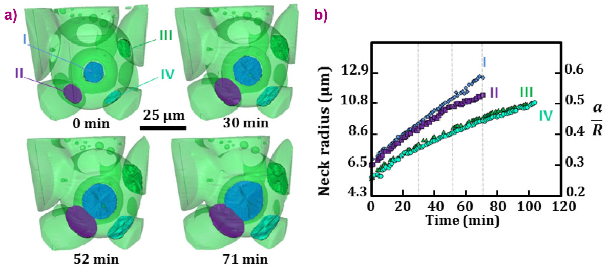
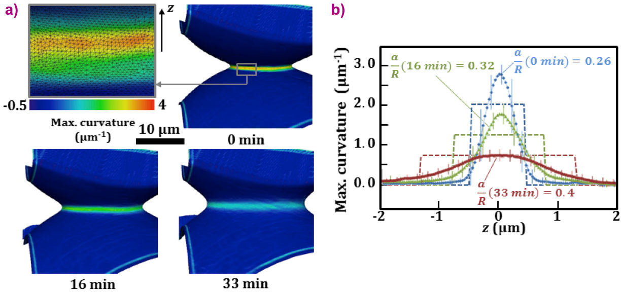
Glass particles, 40 µm in size, were heat treated at 670°C for 105 minutes. During this time period, 90 tomography scans were recorded, each taking 33 seconds and with a pixel size of 100 nm. When compared to the state-of-the-art, this corresponds to a scan time divided by 15 and a pixel size divided by 7. Such high resolution made it possible to follow the formation and growth of micronic necks, as reported in Figure 1. The sizes of four necks around a selected particle were assessed to characterise growth kinetics. The classical Frenkel two-sphere model, which predicts neck size growth scaling with the square root of time, is not supported by our experiment despite being previously supported by in situ 2D observation. This shows that simplistic two spheres models are not applicable to complex 3D configurations of particles. The neck curvature was also measured for a selected neck and is reported in Figure 2 as a function of the distance from the contact. The tangent circle approximation used in the Frenkel model and in many other densification models is not valid here. The neck extension is more limited but with a more pronounced curvature, especially at the very beginning of sintering.
 |
|
Figure 1. a) 3D rendering of the investigated volume showing the growth of four necks (I to IV) during the sintering process at 670°C. b) Evolution of the neck radii with time. a/R refers to the relative neck radius with neck radius: a and particle radius: R. The dashed vertical lines highlight the times when the 3D images in (a) were recorded. |
In summary, 3D in situ observation of particle sintering at high temperature was obtained with an unprecedented combination of high spatial resolution and fast scan time. This experiment allowed the first determination of the local neck curvature, an essential governing parameter for sintering. Our observations lead to the conclusion that existing models, although useful in many cases to predict densification at the macro scale, are based on an invalid assumption (constant neck curvature) and their use is questionable when observing sintering at the local scale. The reported experimental data set the basis for the development of new models. Future experiments target temperatures above 1000°C and even higher resolution. High-temperature X-ray nanotomography should find application in many other fields to reveal previously inaccessible fast phenomena such as nucleation and growth of pores and cracks, glass synthesis, and high-temperature mineralisation processes.
 |
|
Figure 2. a) Evolution of 3D maximum principal curvature rendering of neck IV with time. The first image is an enhanced view of the neck highlighting the surface mesh provided by the high resolution of the experiment. b) Comparison between experimental maximum principal curvature (symbols) and the tangent-circle approximation (dashed lines) as a function of the distance z from the neck plane. |
Principal publication and authors
Fast in situ 3D nanoimaging: a new tool for dynamic characterization in materials science, J. Villanova (a), R. Daudin (b), P. Lhuissier (b), D. Jauffrès (b), S. Lou (b), C.L. Martin (b), S. Labouré (a), R. Tucoulou (b), G. Martínez-Criado (b,c), L. Salvo (b), Materials Today (2017); doi: 10.1016/j.mattod.2017.06.001.
(a) ESRF
(b) University Grenoble Alpes, CNRS, SIMaP, Grenoble (France)
(c) Instituto de Ciencia de Materiales de Madrid, Consejo Superior de Investigaciones Científicas, Cantoblanco (Spain)
References
[1] R.M. German, Sintering: From Empirical Observations to Scientific Principles, Butterworth-Heinemann (2014).
[2] O. Lame et al., Acta Mater. 52, 977–984 (2004).
[3] D. Bernard et al., Acta Mater. 53, 121–128 (2005).
partners


European Synchrotron Radiation Facility - 71, avenue des Martyrs, CS 40220, 38043 Grenoble Cedex 9, France.