- Home
- News
- Spotlight on Science
- Ancient fish illuminates...
Ancient fish illuminates evolution of tooth replacement
13-12-2016
The internal structure of a jaw of the 424 million year old fossil fish Andreolepis has been examined by researchers from Uppsala University and the ESRF using propagation phase contrast synchrotron microtomography (PPC-SRμCT) with sub-micron resolution. Their results reveal that Andreolepis shed its teeth repeatedly by resorption of the tooth bases: this is the earliest known example of a shedding process found in the great majority of bony fishes and land vertebrates, including ourselves.
Share
A tiny jaw of a 424 million year old fossil fish, recently described in Nature, is helping researchers from Uppsala University and the ESRF to understand the evolutionary origin of the shedding of milk teeth in humans. A tooth is shed in the distinctive manner common to all ‘osteichthyans’, the group comprising bony fishes and land animals. The tooth base is resorbed: specialised cells called odontoclasts move in and dissolve the tooth tissue so that the tooth comes loose and drops out of the jaw. This is fundamentally different to the way sharks shed their teeth, which does not involve any resorption of tooth tissue. The two systems seem to have evolved independently from ancestors that kept their teeth throughout life without ever shedding them. Until now little was known about the origin of the osteichthyan tooth shedding system, but this is changing thanks to a fossil fish called Andreolepis and the tomographic capabilities of synchrotron radiation.
During the last few years, we have made extensive use of propagation phase contrast synchrotron microtomography (PPC-SRμCT) at beamline ID19, with voxel sizes below 1 μm, to study the three-dimensional microstructure of fossil bones [1,2]. This makes it possible to retrieve life history data from the bones and to reconstruct their growth histories in detail: sequences of buried structures such as resorption surfaces (where the bone was resorbed, but later regrew, leaving a visible ‘scar’ in the bone) can be modelled and their overlap relationships mapped, allowing the researchers to understand the sequence of steps by which the bones grew.
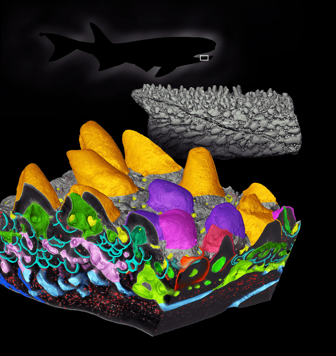
We applied this technique to a jaw bone of Andreolepis, a fossil fish from Sweden that is one of the very earliest osteichthyans. Andreolepis is not known from complete fossils, all the material consists of isolated scales and bone fragments, and this causes problems for the interpretation. In fact, the jaw bone that we studied had been dismissed by some other researchers as not being a jaw at all; for one thing it doesn’t carry a proper tooth row, only a field of tiny tooth-like structures, which might simply be surface lumps and bumps (known as “dermal ornament”) of a kind frequently seen in early vertebrates. However, even though the fossils of Andreolepis are fragmentary, their internal microstructure is perfectly preserved (Figure 1). Underneath the teeth of the jaw lie multiple resorption surfaces: up to four cup-shaped surfaces under each tooth, stacked one on top of another. Each cup represents one occasion when the tooth was shed by basal resorption and then replaced by a new tooth growing in its place.
 |
|
Figure 1. Composite image showing: (back) a silhouette of a generic early bony fish, representing Andreolepis, with a white box indicating the position of the jaw fragment; (middle) the jaw fragment, rendered from a low-resolution PPC-SRμCT scan; (front) a box model of part of the biting surface of the jaw fragment, rendered from a high-resolution PPC-SRμCT scan. Gold: teeth; purple: dermal ornament; green: pulp cavitites; turquoise: buried resorption surfaces; small red spots: bone cell lacuane. The length of the jaw fragment is approximately 6 mm. |
At a stroke, this settled the debate about the identity of the jaw. Dermal ornament is never shed and replaced, so the tooth-like structures are definitely true teeth. Furthermore, only osteichthyans shed and replace their teeth in this way. Andreolepis is, by a wide margin, the earliest osteichthyan in which the process of tooth replacement can be studied. The process seems to have worked in a similar way to primitive modern bony fish, where the replacement tooth grows alongside the old tooth, rather than underneath it as in our own jaws. However, where modern fishes have a well-organised tooth row with a strict pattern of tooth replacement (for example, every other tooth in the row is replaced at the same time in some fishes), in Andreolepis each tooth in the tooth field seems to have had its own replacement cycle. This probably represents the ultimate ancestral condition for all osteichthyans including ourselves.
The unique capabilities of PPC-SRμCT at the ESRF permit the investigation of early vertebrate microstructures revealing their internal three-dimensional architecture. These structures are not only informative about the interrelationships and evolution of early vertebrates: they also give us unprecedented insight into their lives.
Principal publication and authors
The stem osteichthyan Andreolepis and the origin of tooth replacement, D. Chen (a), H. Blom (a), S. Sanchez (a,b), P. Tafforeau (b), P.E. Ahlberg (a), Nature 539, 237-241 (2016); doi: 10.1038/nature19812.
(a) Uppsala University, Uppsala (Sweden)
(b) ESRF, Grenoble (France)
References
[1] S. Sanchez et al., Microscopy and Microanalysis 18, 1095-1105 (2012).
[2] S. Sanchez et al., Nature 537, 408-411 (2016).
partners


European Synchrotron Radiation Facility - 71, avenue des Martyrs, CS 40220, 38043 Grenoble Cedex 9, France.