- Home
- News
- General News
- Scientists uncover...
Scientists uncover how the spine's smallest structures help discs withstand everyday loads
04-11-2026
New synchrotron imaging technology reveals how collagen fibres and molecules work together to absorb and distribute forces inside spinal discs, offering new insight into the underlying disc degeneration, a major contributor to back pain. The study is published in Nature.
Share
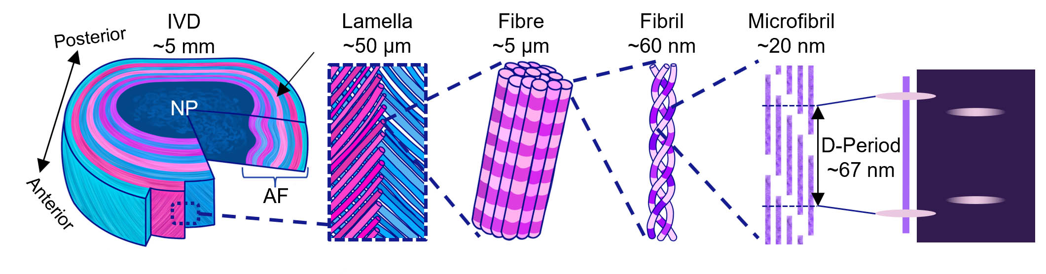
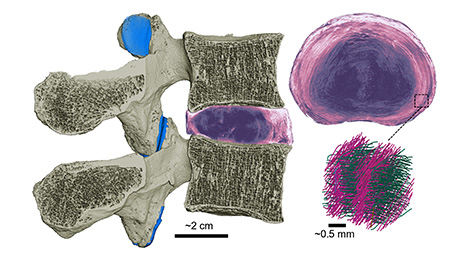
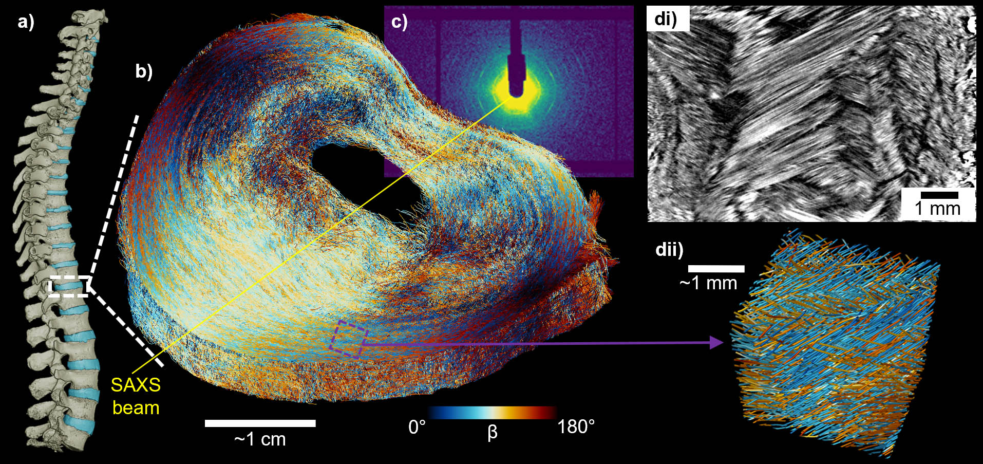
Scientists at Queen Mary University of London and University College London (UCL), in collaboration with the ESRF -the European Synchrotron and Diamond Light Source, have developed a new way to see how the spine’s intervertebral discs respond to mechanical forces, from the scale of the whole tissue down to individual collagen molecules (Fig 2). The technique called TomoSAXS is a new 3D X-ray imaging approach that combines two synchrotron techniques.
 |
|
Figure 1: Structural hierarchy of the intervertebral disc (IVD), from the whole disc (left) to the nanoscale organisation of its collagen fibres and molecules (right). |
The findings could help researchers better understand why spinal discs can withstand the repeated stresses of everyday movement, while also becoming vulnerable to damage and degeneration.
Dr. Alissa Parmenter, joint first author, explains: “When we bend or lift, the forces within our spinal discs are shared between structures far smaller than we can see. With TomoSAXS, we can follow how the tissue deforms and connect that movement to changes within its nanoscale collagen building blocks. This helps us understand how the disc’s intricate organisation allows it to carry load, and why some regions may be more vulnerable to damage.”
 |
|
Figure 2: Tomographic rendering of a spinal segment showing vertebrae compressing the intervertebral disc (IVD) (left), a disc cross-section (top right), and magnified collagen fibres within the IVD (bottom right) |
Despite their importance, scientists have struggled to understand exactly how the different structures inside a disc work together to manage mechanical loads.
 |
|
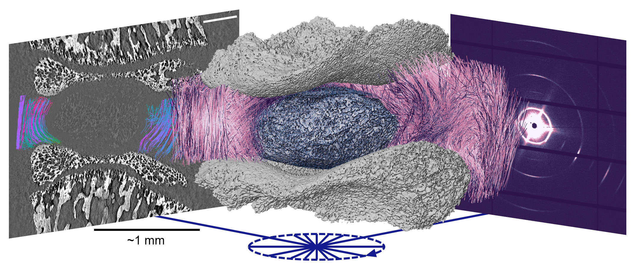
Figure 3: TomoSAXS: imaging the spine across scales. Combining X-ray tomography (left) with small-angle X-ray scattering (right) reveals the three-dimensional organisation of collagen within the intervertebral disc (centre), from the microscale down to the nanoscale. |
For this reason, the research team has developed TomoSAXS, a new 3D X-ray imaging approach that combines two synchrotron techniques to map the structure and mechanics of the disc across multiple length scales – from individual collagen fibrils, which are tens of nanometres across, through to fibres in intact intervertebral discs (IVDs) (Fig 3). These measurements rely on the extremely bright X-ray beams produced by synchrotrons. Using intact IVDs and adjoining bone under compression, the researchers found that the mechanical behaviour of collagen fibres is closely linked to what happens within their constituent fibrils.
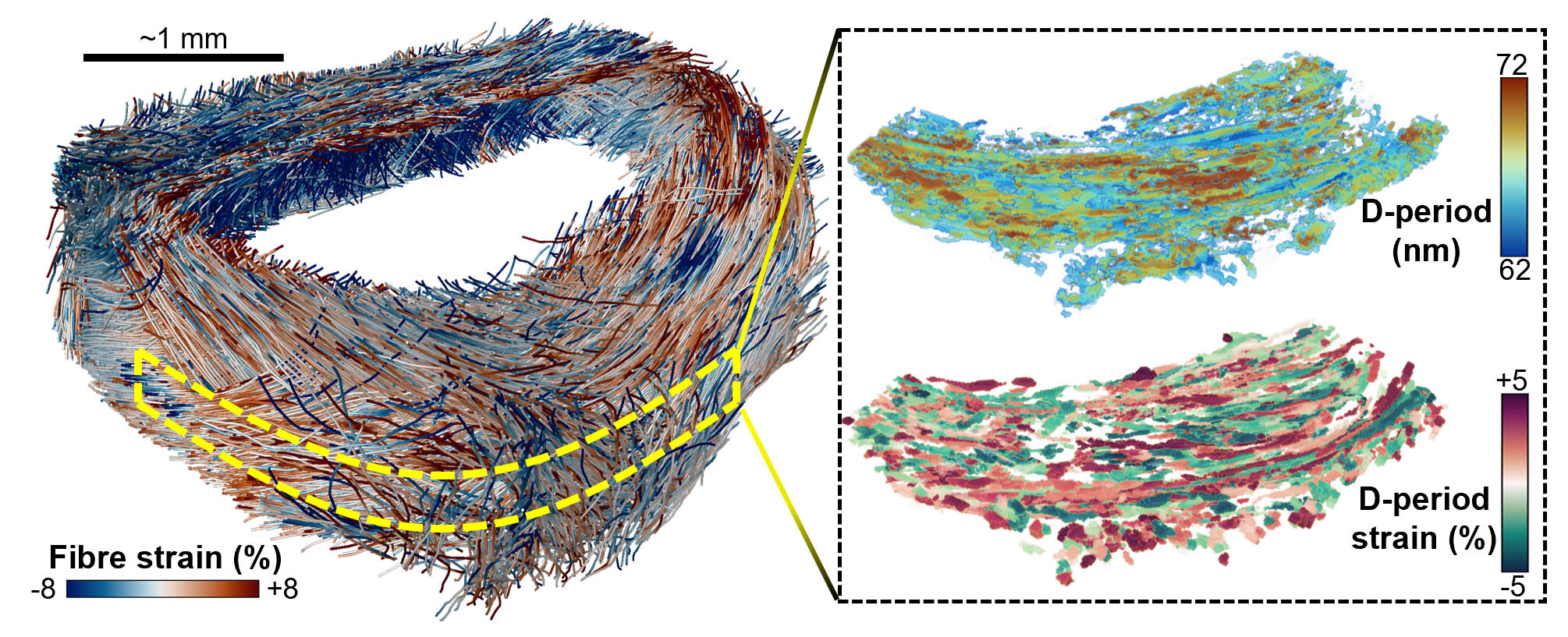
The study shows that collagen fibrils are already under pre-strain before compressive forces are applied during motion, limiting damage. This pre-strain varies across the layered structure of the disc and is closely related to how individual fibres respond to loading (Fig 4).
 |
|
Figure 4: TomoSAXS reveals how collagen fibres are arranged and strained within the intervertebral disc (IVD) (left), and how their nanoscale structure and strain vary (right). |
This suggests that the disc's ability to withstand everyday forces depends on a highly coordinated hierarchy, with structures at the molecular, nanoscale and microscale working together.
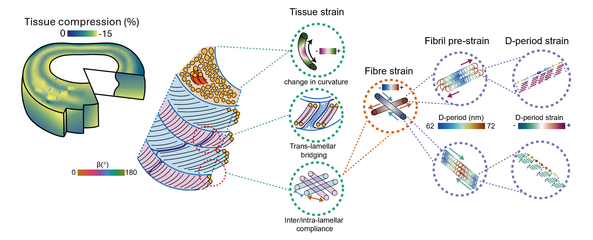
The researchers also found that highly curved collagen fibres experience greater strain. These highly curved regions were particularly common around the transition between the disc’s inner and outer regions, potentially helping to explain why some areas of the disc are more vulnerable to damage and herniation (Fig 5).
Professor Himadri Gupta, joint senior author, from Queen Mary University of London, explains: “The findings point to a potential trade-off in the way the disc is built. We found that substantial pre-stress within collagen fibrils may help the disc achieve the stiffness it needs to perform its role but could also leave some structures with less capacity to withstand additional damaging loads.”
 |
|
Figure 5: How the intervertebral disc (IVD) carries loads, from compression of the whole disc to nanoscale changes in collagen, as revealed by TomoSAXS. |
In simple terms, the same mechanical properties that help the disc do its job may also contribute to its vulnerability over time.
A study based on ESRF’s unique capabilities
After studying small-animal discs under load at Diamond Light Source, the team demonstrated at the ESRF that TomoSAXS can be used to study at multiple scales an intact human intervertebral disc around 5 cm across, an order of magnitude larger than samples previously studied with 3D X-ray scattering methods (Fig 6).
 |
|
Figure 6: TomoSAXS can image whole human intervertebral discs (IVDs), revealing the 3D arrangement of collagen fibres throughout the disc (centre) and their detailed organisation at higher magnification (right). |
“This was only possible at ESRF-The European Synchrotron, where high energy and coherent X-rays enabled tomographic imaging on BM18 of human IVDs with micron resolution and small angle X-ray scattering (SAXS) on ID31. TomoSAXS then enabled matching sub-nanometre displacements in fibril d-spacing with fibres in intact human IVDs (over 5cm in diameter), spanning almost 8 orders of magnitude in strain resolution.” States Prof. Peter Lee (UCL), joint senior author.
“Understanding how damage develops in the intervertebral disc requires us to look beyond the tissue as a whole. With this approach, we can start to connect what is happening to individual collagen structures with how the disc behaves as an organ. This could give us new ways to investigate the mechanical changes associated with ageing and disease.” adds Dr Elis Newham (joint first author).
 |
|
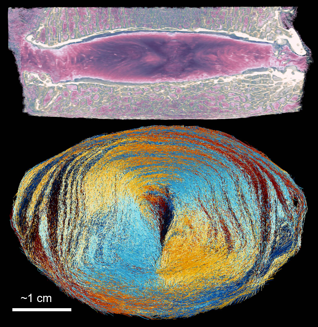
Figure 7: Degenerated human intervertebral disc (IVD), where TomoSAXS could reveal how degeneration alters collagen structure from fibres down to nanoscale fibrils. |
On beamline ESRF’s BM18, the team used Hierarchical Phase-Contrast Tomography (HiP-CT) to image the whole disc at 10 micrometres resolution and trace its collagen fibres through the entire organ. On beamline ID31, high-energy X-rays (75 keV) able to cross several centimetres of tissue recorded scattering patterns through the disc at 24 angles. Combined, the two datasets produced a map of nanoscale collagen properties across a full section of the human disc.
« Integrated X-ray characterization workflows, such as this work, are a major leap towards the understanding of biological and other complex systems. It’s a real gamechanger." Says Jakub Drnec, beamline responsible at ESRF’s ID31.
“It’s the first time an organ was probed at beamline ID31, which is usually dedicated to materials science. Prof. Gupta and his team needed ID31’s high energy and flux as they were measuring scattering from large biological samples (>5cm) with sub-nanometre resolution of strains. Although the experiment was highly ambitious it leads to scientifically relevant results, proving the high versatility of ID31. I hope it will pave the way for other project of this kind.” adds Valentin Vinci, ESRF Scientist at ID31.
At the ESRF, this work extends the Human Organ Atlas project, which uses the innovative HiP-CT technique to image intact human organs in three dimensions down to the cellular scale. Funded in part by the Chan Zuckerberg Initiative, this study is a step towards turning the Atlas from a static database of organ structure into a dynamic one that captures how organs behave under load. "With the Human Organ Atlas, we have been developing ways to visualise human organs in unprecedented three-dimensional detail. TomoSAXS adds another dimension to this approach: rather than only seeing the structure of an organ, we can begin to understand how that structure behaves mechanically, connecting what happens at the scale of the whole organ with changes occurring in collagen at the nanoscale." explains Joseph Brunet, researcher at UCL and ESRF visiting scientist.
The researchers stress that the work does not provide a new treatment for back pain or disc degeneration. Instead, it provides a new experimental framework for understanding the underlying mechanics of healthy and diseased spinal tissue.
In the future, TomoSAXS could be used to investigate how the structure and mechanical behaviour of intervertebral discs change with ageing and disease (Fig 7), and to evaluate potential therapies or new approaches to repairing or replacing damaged discs.
The technique could also be applied to other fibrous biological tissues, including tendons, cartilage and arteries, as well as engineered materials such as carbon-fibre composites.
By connecting structures that range from individual molecules to whole organs, the researchers hope the approach will help uncover where mechanical damage begins and how it develops – providing a stronger foundation for future research into preventing and treating musculoskeletal disease.
Reference:
Newham, E. et al, Nature, print version Nov 5, 2026. DOI: https://doi.org/10.1038/s41586-026-11148-5
partners


European Synchrotron Radiation Facility - 71, avenue des Martyrs, CS 40220, 38043 Grenoble Cedex 9, France.