- Home
- News
- General News
- Regenerating the...
Regenerating the enthesis fascicles with biomimetic electrospun nanofibrous metamaterials
03-10-2025
Researchers have developed an innovative auxetic scaffold that replicates the tendon/ligament-to-bone fascicle organisation of collagen fibers. This could aid in the regeneration of damaged enthesis (spot where the tendon or ligament attaches to the bone). They used the ESRF to test scaffold’s biomimetic deformation under tensile load. The results are out now in Advanced Functional Materials.
Share
You may not have heard the word enthesis, but it is an area in our bodies that can often be injured in high-impact sports and in daily life. The enthesis is the point where tendons or ligaments attach to bone, a vulnerable connection that can be damaged suddenly in accidents or gradually through repetitive micro-injuries over time. Such damage often leads to chronic pain, and current treatment options remain limited because the bone is stiffer than the tendon creating a mechanical mismatch.
Now scientists led by the MERLN Institute for Technology-Inspired Regenerative Medicine at Maastricht University have created a scaffold designed to copy an enthesis fascicle (natural micrometric bundle-like hierarchical level of organisation in tendons or ligaments), capturing its region-specific collagen fibrils’ organization. In fact, the enthesis accounts for the presence of three different tissues organized in series: the tendon/ligament, the non-mineralized fibrocartilage and the mineralized fibrocartilage. “Our aim was to design a material imitating nature so that ultimately it could be used to promote the regeneration of enthesis, which is currently quite challenging, replicating also the typical auxetic effect of the non-mineralized fibrocartilage”, explains Alberto Sensini, one of the principal investigators of the study.
The results show that the scaffold, which was developed using electrospun nanofibers made of a blend of poly-L-lactic acid and collagen, replicates all the orientations/dimensions of collagen fibrils (hundreds of nanometres in size) in the different regions of enthesis (tendon/ligament, non-mineralized and mineralised fibrocartilage). It also reproduces the different region-specific dimensions of a typical enthesis fascicle (from 300 to 600 micrometres), as well as the conical shape of the non-mineralised fibrocartilage.
The scaffold guided stem cells to grow into tendon/ligament-, cartilage-, and bone-like tissue, with each region producing the right supportive matrix thanks to the scaffold’s built-in stiffness differences, and this was better when no extra minerals were added. In mechanical tests, the scaffold closely reproduced how the natural tendon/ligament-to-bone junction behaves, with its conical design allowing smooth transitions between regions.
Strong collaborative effort
The team then came to the ESRF’s ID19 and ID16B beamline to use X-ray imaging techniques with the aim of testing how much the scaffold mimics the multiscale mechanics of natural tendon-bone junction. “We designed these blue-sky biomimetic scaffolds to express a similar auxetic behaviour of an enthesis fascicle; basically, when a tensile load is applied to this tissue, its typical conical shape of the non-mineralized fibrocartilage region expands themselves (i.e. auxetic behaviour) reducing the stress concentrations produced during the passage from the soft tendon/ligament to the stiff bone, increasing the fascicle mechanical stability”, says Professor Lorenzo Moroni, another of the principal investigators. “But we needed experimental proof of that, and that is where the ESRF came in thanks also the extensive expertise of Professor Gianluca Tozzi (University of Greenwich, United Kingdom) in synchrotron tomography and digital volume correlation of biomaterials, which was pivotal in guiding the characterisation activities at ESRF”, he adds.
 |
|
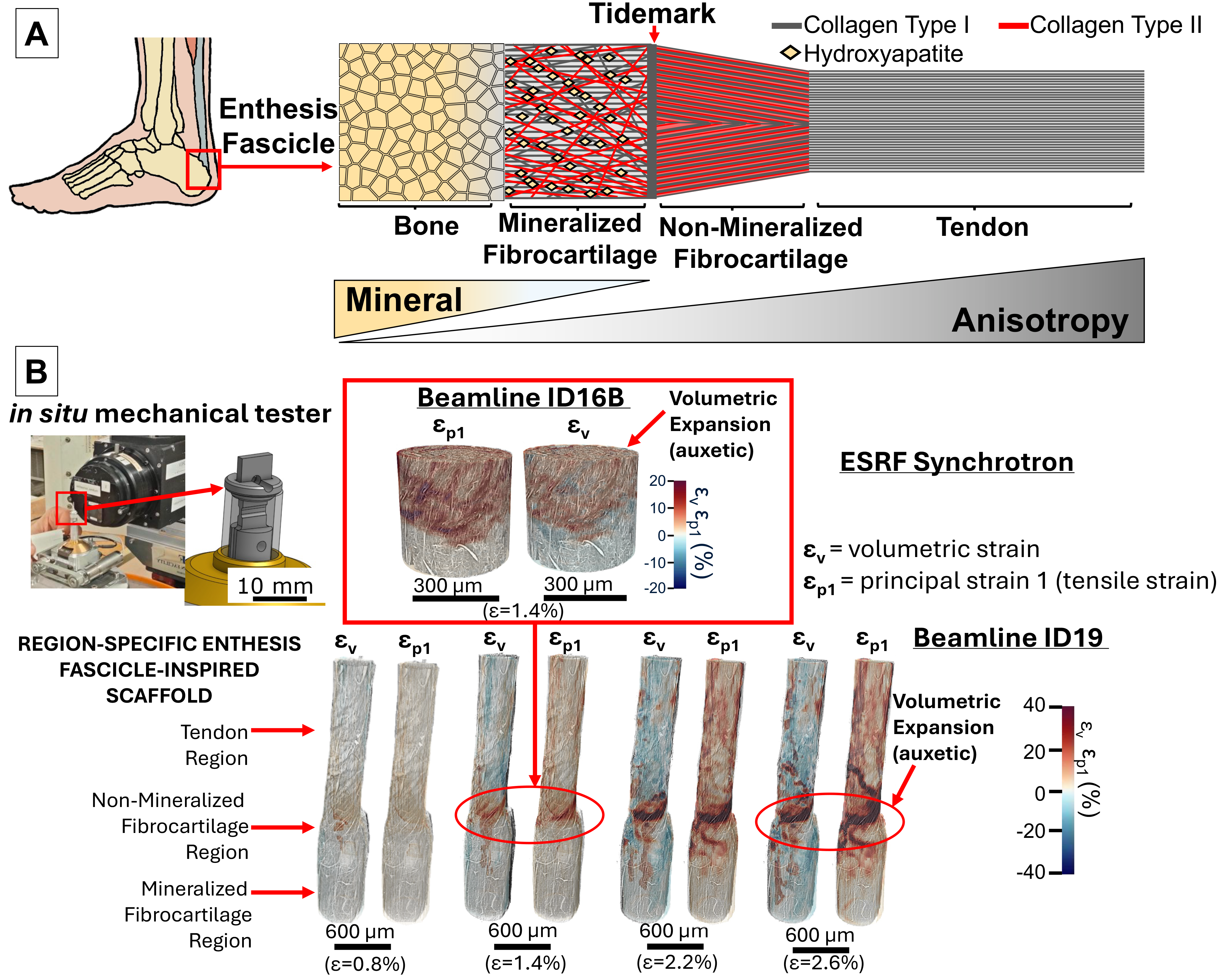
A) Structure and composition of the Extracellular Matrix (ECM) of a natural tendon-to-bone (enthesis) fascicle. B) Results of the multiscale in situ mathematical tensile stepwise test of the electrospun enthesis fascicle-inspired scaffolds at the ID19 (micro x-ray tomographies) and ID16B (nano x-ray tomographies) beamlines of ESRF. The digital volume correlation analysis performed highlights, in response of a tensile strain of the scaffold (principal strain 1), a volumetric expansion (volumetric strain) of the conical non-mineralized junction region of scaffolds mimicking the natural enthesis fascicles (red = tensile train/volumetric expansion; blue = compressive strain/volumetric contraction). |
Thanks to the capabilities of the ESRF, the researchers investigated the multiscale full-field strain distribution of scaffolds, correlating high-resolution x-ray tomographies (with a nanometric voxel size) by using digital volume correlation, during an in situ tensile stepwise test. “The success of this experiment reflects a strong collaborative effort. Despite significant experimental challenges, multi-scale measurements between ID19 and ID16B were achieved using the tensile device developed at the Centre des Matériaux, within the framework of a Long-Term proposal at ID11, together with the SPAM software for 3D deformation field retrieval. This collaboration provided a unique opportunity to apply advanced engineering tools to biomimetic research” says Julie Villanova.
First proof-of-concept
This scaffold is the first proof-of-concept of a nanofibrous device, made by using the electrospinning technique, able to show biomimetic and region-specific auxetic mechanical properties supporting stem cells proliferation/differentiation.
“These findings provide a strong foundation for developing future medical treatments for the enthesis regeneration driven by electrospun metamaterials. On top of that, the digital volume correlation study performed is the first, worldwide, able to describe and document the full-field strain distribution of electrospun nanofibers at the nanoscale during a mechanical test, furnishing a remarkable step forward toward the field of electrospun nanofibrous materials and their mechanics”, concludes Professor Martijn van Griensven, also corresponding author of the publication.
This work is part of the Horizon Europe Marie Skłodowska-Curie postdoctoral Fellowship project 3NTHESES (n. 101061826).
Reference:
Sensini, A. et al., Adv. Funct. Mater. (2025): e11660. https://doi.org/10.1002/adfm.202511660
Text by Montserrat Capellas Espuny
Top image: Zoom-in at the conical non-mineralized junction region of the electrospun enthesis fascicle-inspired scaffold during the in-situ tensile test at ID16B. The two components of strain calculated via Digital Volume Correlation highlights during the tensile stretching of the scaffold (tensile strain) an auxetic volumetric expansion of the conical region (volumetric strain).
partners


European Synchrotron Radiation Facility - 71, avenue des Martyrs, CS 40220, 38043 Grenoble Cedex 9, France.