- Home
- News
- General News
- Novel injector allows...
Novel injector allows X-rays to map membrane proteins
06-05-2014
In the last few days the ESRF’s ID13 beamline has been a hub of activity as it welcomes the largest team of users in its 20-year history. This international team of scientists from the USA, Germany, Switzerland, the UK, France and Sweden are testing a novel lipid cubic phase (LCP) jet injector in the hope of opening new paths into the investigation of membrane proteins.
Membrane proteins, which include G-protein-coupled receptors, are responsible for many vital functions of the cells and play a key role in human health. They are a prime target for developing drugs to treat diseases such as hypertension, asthma, Parkinson’s disease, Alzheimer’s and schizophrenia. Today, more than 50% of all drug targets are membrane proteins.
However, these proteins are notoriously difficult to crystallize. Even when crystals can be obtained, they are very small, diffract badly, are sensitive to radiation and badly withstand being frozen. Obtaining larger crystals is a costly and time-consuming enterprise.
The new injector developed at the Arizona State University by an international team led by Uwe Weierstall aims at investigating the very small and easily produced crystals that cannot be studied today using conventional techniques.
The scientists fed a stream of crystals of the membrane protein bacteriorhodopsin into the path of the X-ray beam using the novel lipid cubic phase (LCP) jet injector. This steady stream of crystals increases the possibility of obtaining a hit, or a diffraction pattern as the pulsed X-ray beam coincides with the sample crossing its path. The crystals are injected at room temperature in an ambient or vacuum environment , just two further advantages of this new sample environment.
This slow motion sequence shows the series of diffraction patterns obtained on a bacteriorhodopsin crystal as it is is subject to high intensity X-rays on the ESRF's ID13 beamline. Credit: U. Weierstall, Arizona State University
Tens of thousands of crystals are being fed through the beam over the 4 days of beamtime. That’s just 96 hours of research time and the sixteen-strong team is making sure they will not miss a second of it! Several crews are working in shifts to ensure the sample environment, data collection and processing, injection and synchrotron instrumentation.
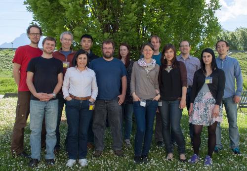
Members of the day crew outside ID13 experimental hutch at the ESRF. Credit: ESRF/C. Argoud
Isabel De Moraes from the Imperial College London and the Diamond Light Source in the UK, wasn’t expecting the experience to be so fulfilling and conclusive: “It’s been an amazing experience to see all these people from different horizons working together, so engaged and focussed on the science taking place.” Isabel was charged with the scientific coordination of the team during beamtime at the ESRF. “Everyone is so highly motivated and involved in their work, there is almost no need for coordination!”
“We came to the ESRF expecting to test the crystals to see if they would diffract, however the results have been so amazing that we are in the process of collecting a complete data set to see if we can solve a structure!” says Uwe Weierstall. “The beamline staff at ID13 have worked extremely hard to prepare for our experiment and make sure that all runs smoothly while we are here. Really, all aspects of our experience here have surpassed expectations.”
The injector was originally designed for use at Free Electron Laser facilities which have a higher photon flux allowing them to reduce exposure time and map small crystals before they are damaged.
Later this year ID13 will be equipped with a detector that will increase the frame rate from 17Hz to 700 Hz opening new possibilities for clearer diffraction patterns. Along with the expected upgrade of the accelerator complex in 2018-2019, the higher photon flux will maintain the ESRF as a complementary facility for serial crystallography alongside Free Electron Laser facilities.
This international collaboration brings together scientists from the Arizona State University (USA), the Scripps Research Institute (USA), the Paul Scherrer Institute (CH), Desy/Petra/C-FEL (D), Diamond Light Source (UK), Imperial College London (UK), Gothenburg University (S) and the European Synchrotron, ESRF (F). Financing of the project was obtained through NanoMem-EU project coordinated by Richard Neutze from Gothenburg University, the main PI of the project.
Find out more about the ESRF's ID13 beamline.
Top image: Uwe Weierstal surveys the injection process from the experimental hutch of ID13. Credit: ESRF/C. Argoud
partners


European Synchrotron Radiation Facility - 71, avenue des Martyrs, CS 40220, 38043 Grenoble Cedex 9, France.