- Home
- News
- General News
- New insights into...
New insights into bone structures shake up current models
10-06-2020
Bones may not have the structure that was previously believed, an international team of scientists have found using newly developed X-ray tensor tomography.
Share
Bone tissue is a highly specialized tissue, with a structure tailored to the specific functions of the body. A healthy bone is both strong, with great bearing capacity, and tough, with great breaking strength. An increased understanding of the basic structure of bones could lead to the prevention of various bone diseases and the development of completely new materials with unprecedented properties.
An international team of scientists from Aarhus University (Denmark), the European Synchrotron, Chalmers University (Sweden) and the Paul Scherrer Institute (Switzerland) has found a new substructure in the bone tissue using the new method of x-ray tensor tomography on beamline ID13 at the ESRF. The findings open up new approaches to study the underlying architecture of bone tissue and create a better understanding of biological materials.
The healthy architecture of bone tissue is made up of basically two types of building blocks: the so-called collagen fibrils, which primarily consist of the protein collagen, and the calcium phosphate nanocrystals, which weave the fibrils together.
Jointly, the two types of tissue form a coherent hierarchical structure that combines the ability of the fibrils to bend with the toughness, hardness and resistance of the nanocrystals. It is this twisted structure that gives the bones their mechanical properties, but researchers still do not fully understand it. “The challenge during all these years was that there was no method to detect the orientation of nanocrystals in the bone tissue”, explains Henrik Birkedal, scientist at Aarhus University and corresponding author of the publication.
The team at the ESRF’s ID13 beamline joined forces with the Birkedal group from Aarhus University and Marianne Liebi from Chalmers University to improve significantly the technique of X-ray tensor tomography, which until now could only allow the study of the nanostructure. Thanks to their efforts, they can now map crystals and nanostructures: it makes it possible to see the way the nanocrystals actually lie in the structure. With the new technique, the researchers have discovered that the lime crystals may not only be homogeneously oriented relative to the fibrils, as previously thought. According to Manfred Burghammer, scientist in charge of ID13: "Our new approach is made possible by the great progress currently being made in the international X-ray synchrotrons, whose power is drastically increasing in these years".
"We were honestly a little bit shocked to find the deviation from the models in what we could see with this new method," says Birkedal. "An interdisciplinary international collaboration has been crucial to our success in making this breakthrough that combines knowledge from different fields, ranging from physics and chemistry to medicine."
 |
|
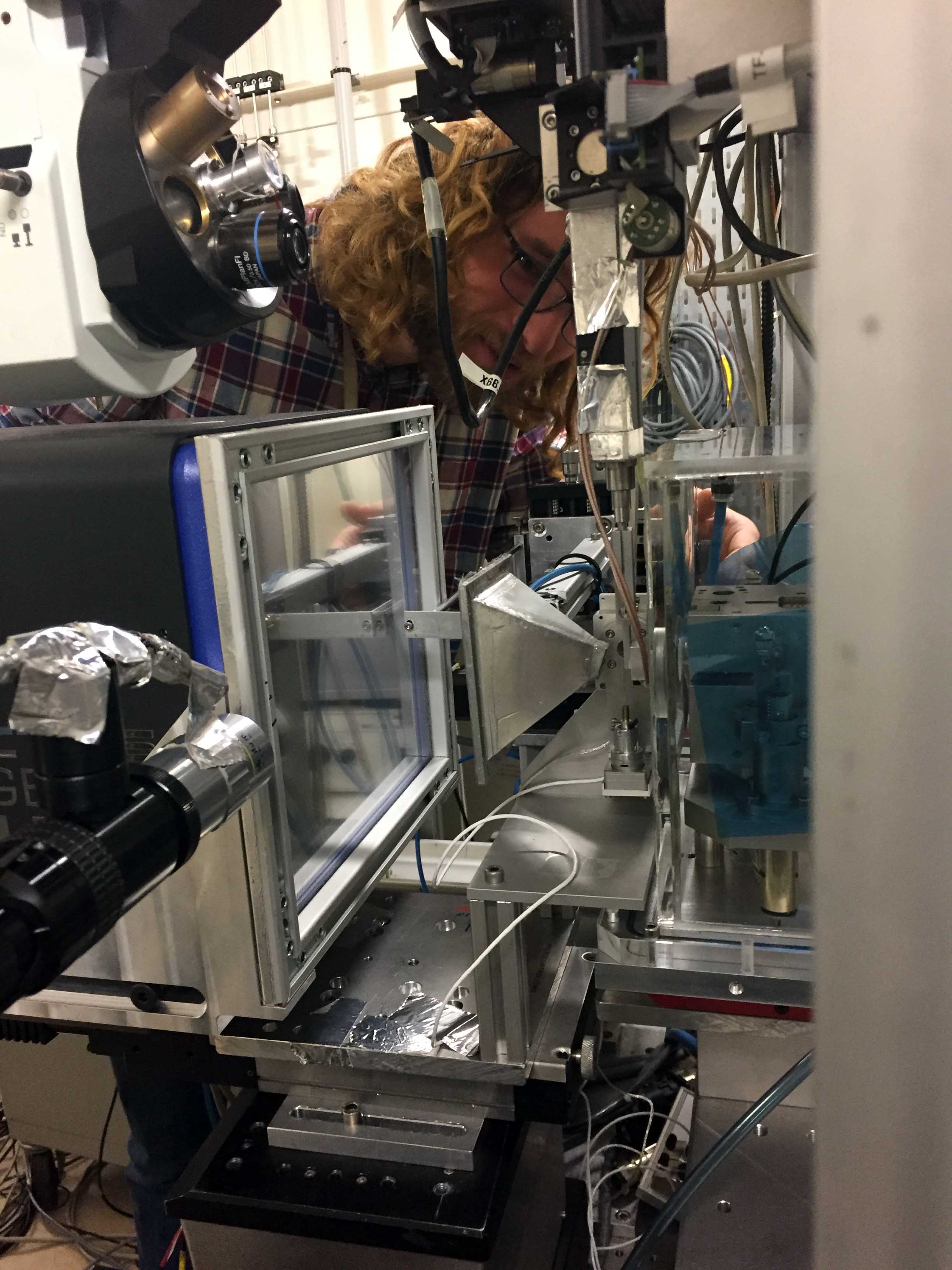
Tilman Grünewald, first author of the paper, mounts the sample on the beamline ID13 at the ESRF. |
The new 3D images surprised the research leaders because it shakes up fundamental theories that state that bones are built in a predominantly uniform hierarchical structure. The finding now raises fundamental questions about some models that have been used to describe, among other things, the bone formation process. “Admittedly, it is premature to give a clear explanation of what lies behind the deviation that has now been detected, but it has given science a new method of looking into the underlying structure of bones, and it shows that important puzzle pieces need to be added to our understanding of bone”, says Tillman Grünewald, former ESRF scientist and currently at the Institut Fresnel, Marseille (France).
“Bones, and other biological materials such as seashells, have mechanical properties that are closely linked to their structure. The better we understand it, the closer we get to being able to emulate nature's construction methods. Our study here has given us a new tool to unveil a few more of nature’s secrets”, concludes Birkedal.
Reference:
Grünewald, T., et al, Science Advances, 12 June 2020. https://doi.org/10.1126/sciadv.aba4171
Top image: The alignment difference between the orientation of the nanostructure of the bones and the oriention of the crystal structure. Credits: Grünewald, T., et al, Science Advances, 2020.
partners


European Synchrotron Radiation Facility - 71, avenue des Martyrs, CS 40220, 38043 Grenoble Cedex 9, France.