- Home
- News
- General News
- Mind the gap - ESRF...
Mind the gap - ESRF tracks defects triggered by composites in root fillings
21-12-2021
Polymer composite fillings of root-canal treated teeth can fail over time. Scientists led by the Charité University in Berlin (Germany) have found that this is not because of the dentist’s lack of skills but rather because of stresses that build up and deform the biomaterial just after it is placed. The results are published in Acta Biomaterialia.
Share
It is one of the most peculiar images that can come to mind: a dentist restoring severely destroyed teeth and placing fillings on a beamline at a synchrotron. It is, however, exactly what happened on beamline ID19 a while back, when a team from the Charité and TU Universities in Berlin and the ESRF examined how well composite fillings adapt to cavities in the tooth root canal orifice.
To treat cavities in teeth, dentists expose solid tooth tissue prior to "filling" the volume of missing structure with rigid biomaterials that sustain chewing forces. In the past, dentists used metals such as amalgam or gold, but today they mostly use composite materials, made of polymer and glass. Such materials, which are well resistant to damage and highly aesthetic, allow rapid recovery of tooth function. However, composites tend to fail in the long run, especially in root-canal filled teeth.
“Until now people believed that when composite fillings failed it was because of an unskilled dentist or because of poor materials, leading to wear and tear. What we have found now is that polymerization stresses created during placement may radically determine how resilient a filling will be”, explains Paul Zaslansky, a dentist and researcher in the Charité University in Berlin and corresponding author of the paper.
Filling teeth on a beamline
Zaslansky and his colleagues studied the damaged teeth as soon as the filling was placed, by combining X-ray phase contrast enhanced time-lapse radiography, digital image correlation and submicrometer-resolution phase-contrast enhanced microtomography at the ESRF’s ID19 beamline. These experiments revealed how much the material deforms due to composite polymerization. The unique imaging configuration on the beamline made it possible to track, almost in real time, the processes that take place as soon as the composite is placed as a viscous mass, injected into the tooth, where it solidifies. For Kerstin Bitter, a teacher and practicing dentist who carried out the ex-vivo ‘treatment’ on the beamline, it was the first time at the ESRF: “I was very impressed by the rich array of available facilities, a scientific ‘town’ on its own, located in a breathtaking landscape”.
By tracking the dynamics of polymerization, the researchers discovered that early in the process, within a minute or so, the injected material condenses and relaxes freely. Shortly afterwards though, when the viscosity of the polymer interferes with the glass elements of the composite, it increasingly deforms due to internal stresses. This stress is very localised and triggers different effects, including deformation of free surfaces, detachment of the root filling or deformation of the tooth cavity walls. In small cavities, gaps tend to appear, and these may allow bacteria to dive into the tooth, leading to reinfection. “Funnily enough, if you have a big filling, the material has more space to flow than in a narrower cavity, so the bigger restoration is going to generate less stresses than the smaller one, which is contrary to what is usually thought”, explains Zaslansky.
 |
|
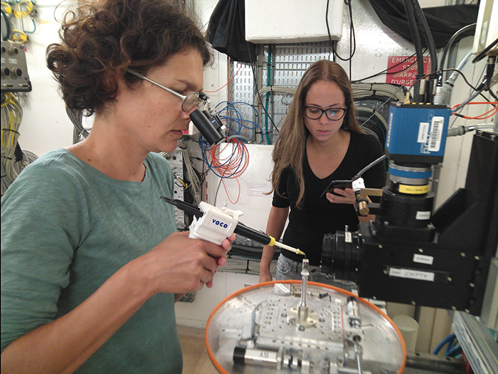
Kerstin Bitter placing a filling on a tooth on ID19's experimental hutch. Credits: K. Bitter. |
“As a dentist, to see in real-time how these fillings delaminate immediately after placement was deeply impressive and a bit shocking”, says Bitter. “It represents a true paradigm shift, as we, dentists, have always believed that we can fully control how these materials behave. The next step is to analyze how these fillings perform over time after simulated loading and to see if these initial gaps progress over time. We also have to analyse if other materials behave similarly and how these initial delaminations can be avoided, for example, by using another filling technique”, she concludes.
Reference:
Bitter, K., et al, Acta Biomaterialia, 2021. https://doi.org/10.1016/j.actbio.2021.10.052
Text by Montserrat Capellas Espuny
Top image: A 3D rendering of the ID19 root-filling data with pores, interface and composite (with particles) in the cavity. Credits: K. Bitter.
partners


European Synchrotron Radiation Facility - 71, avenue des Martyrs, CS 40220, 38043 Grenoble Cedex 9, France.