- Home
- News
- General News
- Inauguration of...
Inauguration of a Cryo-electron microscope platform at the ESRF
10-11-2017
A TITAN KRIOS cryo-electron microscope has been inaugurated at the ESRF, the European Synchrotron, in Grenoble, France. The inauguration took place in the presence of Ada Yonath, chemistry Nobel Prize laureate in 2009, Francesco Sette, Director General of the ESRF and all the partners that jointly run the facility with the ESRF: the European Molecular Biology Laboratory (EMBL), the Institut de Biologie Structurale (IBS) and the Institut Laue-Langevin (ILL). This cryo-electron microscope will provide Europe with a new, innovative and complementary facility for structural biology, serving a vibrant scientific community and addressing new biology and health challenges.
Share
The importance of cryo-electron microscopy (cryo-EM) for biochemistry has been recently recognised through the award of the Nobel Prize in Chemistry 2017 to Jacques Dubochet, Joachim Frank, and Richard Henderson. According to the Nobel Prize Committee “this technique both simplifies and improves the imaging of biomolecules and has taken biochemistry into a new era”. Chemistry laureate Ada Yonath, who received the Nobel in 2009 for determining the structure of the ribosome using X-ray crystallography, in particular at the ESRF, described this technique as “the next step in “the resolution revolution.”
To capture life in atomic details
It is often said in structural biology that “seeing is believing”: It is only by knowing the atom-by-atom arrangement of a biomolecule that researchers can grasp how it works and identify therapeutic solutions.
 |
|
|
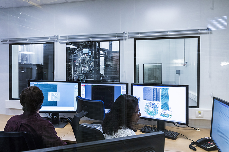
The team in the control hutch of the new CRYO-EM platform. Credits: Stef Candé. |
|
Thanks to cryo-EM, researchers can now freeze biomolecules mid-movement, and therefore portray them at atomic resolution. They can also visualise processes inside cells they have never previously seen or depict membrane proteins. Cryo-EM allows researchers to produce ‘films’ that reveal how proteins interact with other molecules, which is decisive for both the basic understanding of life’s chemistry and for the development of pharmaceuticals. The technique of cryo-EM has opened up the molecular world of the cell to direct observation, with scientists using the technique to probe the structure of drug targets, as well as components within cells involved in sensing pain and human diseases.
A collaborative platform for Europe and beyond
The strength of this equipment lies in its synergy with synchrotron-based techniques and, in its collaborative nature. The cryo-EM platform will provide scientists with a support to users from all the partner institutes: ESRF, EMBL, ILL and IBS.
This platform will offer users in Europe and beyond a holistic approach to their research as it concentrates 350 scientists from three European Institutes and one French mixed research facility in structural biology (MX) on a same site. Along with state-of-the-art and highly automated MX beamlines, it will provide scientists with full services and a large combination of techniques from sample preparation, to data collection and their interpretation.
 |
|
|
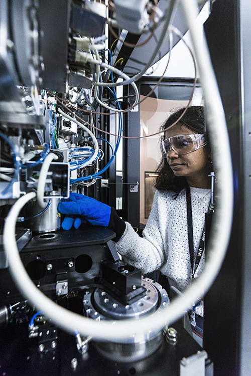
Eaazhisai Kandiah preparing an experiment. Credits: Stef Candé. |
As underlined by Francesco Sette, DG of the ESRF “We are proud to inaugurate this cryo-electron microscope. The ESRF has always helped lead the way in structural biology, with the commissioning of world’s first undulator-based beamlines for MX and MX-dedicated micro-focus beamline. With this collaborative cryo-EM platform, the ESRF, with its partners, will offer a unique hub for pioneering research in structural biology and a great tool for the understanding of life.”
The cryo-EM platform, which started operating in November, is open to the international user community through a peer-review system that evaluates the scientific merit of proposals, as well as their technical feasibility. “There are few cryo-EMs in Europe, and they are mostly used locally. The ESRF, as an international institute, is the ideal place to offer this service to the scientific community worldwide,” explains Eaazhisai Kandiah, beamline scientist.
With the addition of this world-class cryo-EM, the ESRF offers the international structural biology community the possibility of coordinated access to cutting-edge methods and instrumentation. "The ESRF international scientific community can now use the information obtained both from diffraction experiments and from cryo-EM in a complementary fashion to better and more fully interpret action and function of complex bio-macromolecules. It will allow scientists to push the field of structural biology forward, to better understand the nature of health diseases, and to develop new drugs.” underlines Christoph Muller-Dieckmann, beamline responsible.
The Cryo-EM platform will be run by 7 scientists, including 3 from the ESRF, 2 from EMBL and 2 from IBS.
"At EMBL, we're very excited at the opportunities this new facility will offer, and the more refined insight it will give us into detailed biological structures that are difficult to observe otherwise, like the components of viruses," said Iain Mattaj, Director General of EMBL. "We're happy to continue to develop this technology for biology, with an EMBL scientist as one of the platform's managing scientists.”
“The IBS, with its competitive program in electron microscopy, is proud to participate in this European effort to provide state-of-the-art cryo-electron microscopy technology to a large community of structural biologists in order to piece together the atomic details of macromolecular assemblies, the molecular machines of life”, underlined Winfried Weissenhorn, Director of the IBS.
“The arrival of cryo-EM on the EPN campus provides new, state-of-the-art infrastructure and a major boost for biology. The ILL, with its UK partner at Keele University, is excited to be part of this project, looking to further develop synergies between neutron techniques and the wider range of techniques on the EPN campus”, explained Mark Johnson, Director of Research at ILL.
Top image: The team of the CRYO-EM. Credits: Stef Candé.
partners


European Synchrotron Radiation Facility - 71, avenue des Martyrs, CS 40220, 38043 Grenoble Cedex 9, France.