- Home
- Investigating the local structure of a π-π conjugated network by X-ray nanodiffraction and cross correlation
Investigating the local structure of a π-π conjugated network by X-ray nanodiffraction and cross correlation
Poly(3-hexylthiophene) (P3HT) is one of the most widely used semicrystalline polymers in organic electronics. Cast P3HT thin films are typical semicrystalline where well-ordered crystalline nanodomains with sizes of about 5-10 nm are embedded in a disordered host matrix. Considerable effort has been made to understand the charge transport mechanism in terms of the size and orientation of these nanodomains and their degree of crystallinity. It was found that the P3HT films, with edge-on orientation of thiophene moieties with π-π stacking direction along the substrate plane, are a favourable way of achieving high charge mobility in organic field-effect transistor (OFET) devices [1].
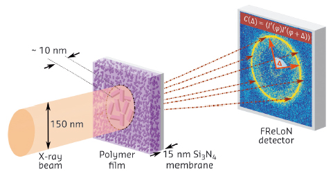
The majority of structural investigations of organic thin films has been performed using the grazing incidence X-ray diffraction (GIXD) technique, where the large footprint accounts for spatial averaging over more than ten thousand crystalline nanodomains. This drawback of GIXD in terms of sample averaging has been overcome by using an X-ray nanobeam in transmission geometry (Figure 43) at the ID13 nanofocus beamline. We employed nanobeam X-ray diffraction using an X-ray spot size of 150 nm to investigate the local structure of P3HT thin films.
 |
|
Fig. 43: Schematic setup of the X-ray scattering experiment employing transmission geometry. |
P3HT drop-cast films on rectangularly shaped grids with 15 nm thick Si3N4 membranes (Dune Sciences, Inc.) were measured by using a mesh scan technique with a step size of 180 nm. From this we obtained 121 scattering patterns.
The (020) diffraction ring associated with the π−π stacking of the thiophenes is the most prominent signal visible at q = 16 nm-1 and originates from the edge-on orientation of the nanocrystals. The broadening of the peaks confirms the size of the nanocrystals of 5−10 nm as deduced by the Debye−Scherrer formula. Due to the small spot size of 150 nm, the X-ray diffraction pattern becomes sensitive to the local crystalline structure and the local angular or orientational order of the crystallites can be traced by the use of X-ray cross-correlation analysis (XCCA) [2].
In comparison to integrated profiles, which do not show any pronounced spatial variation over the whole nanomesh, the correlation functions elucidate the variety of local structures existing in P3HT.
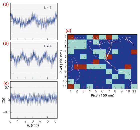
At certain spots, the angular correlations with 2-fold symmetry (Figure 44a) and pronounced 4-fold symmetries (Figure 44b) are found, while other positions show no sign of orientational variation with flat correlation functions C(Δ) (Figure 44c). Despite 2-fold symmetry being a typical motif in the arrangement of P3HT crystals (even on larger length scales), the 4-fold symmetry oscillations provide an enhanced local degree of ordering and cannot be resolved in conventional GIXD measurements.
At each spot on the sample, both the magnitude and the direction (the phase) were deduced for the L = 2 Fourier component of the scattering intensity. The arrows in Figure 44a have length associated with the magnitude of the Fourier component and direction given by the phase. The colour of the pixels represent the magnitude binned into three intervals: strong (brown), medium (green) and weak (blue), respectively.
 |
|
Fig. 44: Correlation functions C(Δ) at four different positions on the sample showing different symmetries: (a) 2-fold, (b) 4-fold, (c) flat correlation at the (020) Bragg peak position q = 16 nm-1, (d) Stream lines (white) of the Fourier component L = 2. |
We observe a strong spatial variation without gradual transition from spot to spot in the correlation functions. The overall appearance of C(Δ) is different at each spot, indicating that the structural motifs are short-range with length scales below the spot size. One key aspect for π−π conjugated polymers is the question of connectivity through the nanoscale complex network. Figure 44d visualises a form of connectivity by displaying streamlines through the different angular order maps. The streamlines indicate how distant parts of the sample can be connected along the orientational order direction. Figure 44d shows that for L = 2 Fourier component it is difficult to connect over long distances when angular disorder is present. The P3HT system possesses a complex interplay between order and disorder, and the understanding of such interconnectivity remains a manifold task.
Our X-ray study reveals the complex nanoscale structure of the crystals in P3HT where spatially resolved investigation provides strong variations of the structural order on a local scale. The diffraction patterns confirm that a high degree of angular order can exist in the polymer network within an X-ray spot size of 150 nm. The XCCA technique allows us to determine local features on the scale of the working device, avoiding the averaging procedures. The present approach investigating the ordering and interconnectivity of the π−π conjugated network at the 150 nm level has particular potential for the optimisation of organic electronic devices.
Principal publication and authors
C. Gutt (a), L. Grodd (a), E. Mikayelyan (a), U. Pietsch (a), R. J. Kline (b) and S. Grigorian (a), J. Phys. Chem. Lett. 5, 2335–2339 (2014).
(a) Fachbereich Physik, Universität Siegen (Germany)
(b) Materials Science and Engineering Division, National Institute of Standards and Technology, Gaithersburg (USA)
References
[1] H. Sirringhaus et al., Nature 401, 685 (1999).
[2] P. Wochner et al., PNAS 106, 11511 (2009).
partners


European Synchrotron Radiation Facility - 71, avenue des Martyrs, CS 40220, 38043 Grenoble Cedex 9, France.