Other materials
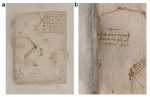
Multi-modal analysis of black stains on the passepartout of Codex Atlanticus Folio 843 by Leonardo da Vinci
XRD micro-analyses (ID13 and ID22, through the historical materials BAG) and µXANES analyses (ID21) have been combined with laboratory imaging techniques to identify the nature of black stains present on the passepartout close to the margins of Folio 843 of Leonardo da Vinci’s Codex Atlanticus. Instead of the microbiological deterioration which was formerly proposed, black nano-particles of metacinnabar (β‑HgS) have been identified as the component of the black staining.
 |
N. Guarnieri, M. Ghirardello, S. Goidanich, D.Comelli, D. Dellasega, M. Cotte, E. Fontana and L. Toniolo, “Imaging and micro‑invasive analyses of black stains on the passepartout of Codex Atlanticus Folio 843 by Leonardo da Vinci”, Scientific Reports, (2023). |
This paper accounts for the diagnostic campaign aimed at understanding the phenomenon of black stains appeared on the passepartout close to the margins of Folio 843 of Leonardo da Vinci’s Codex Atlanticus. Previous studies excluded microbiological deterioration processes. The study is based on a multi‑analytical approach, including non‑invasive imaging measurements of the folio, micro‑imaging and synchrotron spectroscopy investigations of passepartout fragments at different magnifications and spectral ranges. Photoluminescence hyperspectral and lifetime imaging highlighted that black stains are not composed of fluorescent materials. μATR‑FTIR imaging of fragments from the passepartout revealed the presence of a mixture of starch and PVAc glues localized only in the stained areas close to the margin of the folio. FE‑SEM observations showed that the dark stains are localized inside cavities formed among cellulose fibers, where an accumulation of inorganic roundish particles (∅100–200 nm in diameter size), composed of Hg and S, was detected. Finally, by employing synchrotron μXRF, μXANES and HR‑XRD analyses it was possible to identify these particles as metacinnabar (β‑HgS). Further research is needed to assess the chemical process leading to the metacinnabar formation in the controlled conservation condition of Leonardo’s Codex.
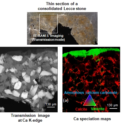
2D XANES mapping to study the consolidation of limestones
Different 2D mapping techniques (µXANES in transmission, in fluorescence and µXRD) have been combined and compared to identify and locate the crystalline an amorphous phases formed during consolidation treatments of limestone.
 |
L. Monico, L. Cartechini, F. Rosi, W. De Nolf, M. Cotte, R. Vivani, C. Maurich and C. Miliani, “Synchrotron radiation Ca K-edge 2D-XANES spectroscopy for studying the stratigraphic distribution of calcium-based consolidants applied in limestones”. Sci Rep 10, 14337 (2020). |
In Heritage Science, the evaluation of stone consolidation treatments by investigating the nature of in situ newly formed products and their penetration depth within the consolidated matrix is a grand challenge. A number of analytical methods have been proposed, but, currently, most of them are not able to supply a full overview of the spatial, structural and compositional information of the newly formed crystalline and amorphous phases with a submicrometric lateral resolution. Here, we examined, the capabilities of synchrotron radiation (SR)-based two-dimensional X-ray absorption near-edge structure (2D-XANES) spectroscopy at Ca K-edge for determining the structural and compositional properties of the compounds formed after the application of a calcium acetoacetate-based consolidant on a porous carbonatic stone (limestone) and for investigating their stratigraphic distribution at the submicrometric scale length. We evaluated advantages and drawbacks of three Ca K-edge 2D-XANES-based approaches: (i) transmission mode full-field-XANES (FF-XANES) imaging; (ii) micro-X-ray fluorescence (μ-XRF) mapping above the Ca K-edge combined with the acquisition of XRF mode μ-XANES spectra at a limited number of spots; (iii) full-spectral µ-XANES (FS µ-XANES) mapping in XRF mode and its variant called selectively induced X-ray emission spectroscopy (SIXES) mapping. Overall, Ca K-edge 2D-XANES spectroscopy provided accurate qualitative and semi-quantitative information on the newly formed calcium carbonates (i.e., amorphous calcium carbonate, vaterite and calcite) and their stratigraphic distribution at the submicrometric scale, thus opening a new scenario to study the carbonatation process of calcium-based consolidants in limestones.
Degradation of organic-based modern materials in artworks
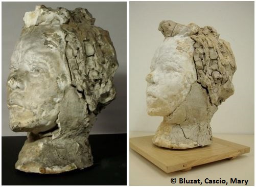
While most of the experiments at ID21 target inorganic materials, in some cases, micro-analyses are carried out on organic-based fragments, usually taken from rather recent (19th-20th C.) objects. As an example, composition and degradation of modern modeling clay in Rodin’s sculptures and of polymers in Italian design objects have been characterized using synchrotron micro-probes.
 |
J. Langlois, G. Mary, H. Bluzat, A. Cascio, N. Balcar, Y. Vandenberghe and M. Cotte, "Analysis and conservation of modern modeling materials found on Auguste Rodin's sculptures", Studies in Conservation, 62, 247-265 (2017). |
Prior to the exhibition Portrait-making, Rodin and his models (2009), the Rodin museum wanted to restore two busts of Hanako and Clemenceau. Interestingly, these two sculptures contain pieces of modern modeling materials (MMMs) invented at the end of the nineteenth century as an alternative to clay or waxes. The poor state of conservation of the two portraits made any handling and exhibition impossible. Accordingly, the purpose of this article is twofold: to contribute to technical art history and conservation. Elemental and chemical analyses were done on samples from 12 sculptures (SEM–EDX, FTIR, GC–MS, GC–FID, XRD, synchrotron-based µXRF, µXANES, and µFTIR) aimed at identifying the composition of MMMs used by Rodin on plaster sculptures and establishing hypotheses about the origins of their degradation. This thorough study of their composition and degradation was necessary to implement an appropriate restoration plan. The development of conservation protocols adapted to such materials is rarely documented. Different tests were performed on mock-ups (pH, solubility, adhesion, consolidation, and cleaning). In particular, a protocol based on laser cleaning was developed and successfully applied to remove superficial dust and crusts so that the sculptures regained their original aspect.
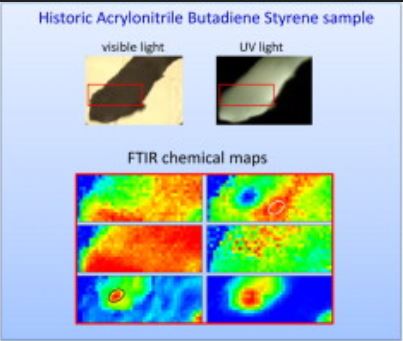 |
D. Saviello, E. Pouyet, L. Toniolo, M. Cotte and A. Nevin, "Synchrotron-based FTIR microspectroscopy for the mapping of photo-oxidation and additives in acrylonitrile–butadiene–styrene model samples and historical objects", Analytica Chimica Acta, 843, 59-72 (2014). |
Synchrotron-based Fourier transform infrared micro-spectroscopy (SR-μFTIR) was used to map photo-oxidative degradation of acrylonitrile–butadiene–styrene (ABS) and to investigate the presence and the migration of additives in historical samples from important Italian design objects. High resolution (3 × 3 μm2) molecular maps were obtained by FTIR microspectroscopy in transmission mode, using a new method for the preparation of polymer thin sections. The depth of photo-oxidation in samples was evaluated and accompanied by the formation of ketones, aldehydes, esters, and unsaturated carbonyl compounds. This study demonstrates selective surface oxidation and a probable passivation of material against further degradation. In polymer fragments from design objects made of ABS from the 1960s, UV-stabilizers were detected and mapped, and microscopic inclusions of proteinaceous material were identified and mapped for the first time.
partners


European Synchrotron Radiation Facility - 71, avenue des Martyrs, CS 40220, 38043 Grenoble Cedex 9, France.