- Home
- Angular absorption dependence at the calcium K-edge reveals bone mineral orientation
Angular absorption dependence at the calcium K-edge reveals bone mineral orientation
A fast and straightforward method to map apatite orientation in human bone tissue is proposed that exploits the sensitivity of X-ray absorption spectroscopy at the calcium K-edge to the apatite crystal orientations.
The scanning X-ray microscope at beamline ID21 is dedicated to micro X-ray fluorescence (µXRF) and micro X-ray absorption spectroscopy (µXANES) in the tender X-ray domain (2.0-9.2 keV). The instrument offers element sensitivity in the low ppm range and localisation of various elements with a sub-micrometric beam. X-ray absorption spectroscopy can be performed in both scanning and full-field (FF) modalities, mainly in XRF and transmission modes, respectively. FF-XANES consists of acquiring radiographic scans of thin samples, illuminating samples with an unfocussed millimetric beam, at X-ray energies around the edge of interest. In this way, up to 106 XANES spectra can be acquired for 2D regions with sub-micron pixel-size in a single stack of radiographs, and in a relatively short acquisition time (here 40 minutes per sample). In this study, XANES spectroscopy in both FF transmission and XRF scanning mode at the Ca K-edge was applied to the analysis of human bone tissue in different pathological states and of varying anatomical origin.
Bone is a hierarchically-structured fibre composite consisting of collagen fibres reinforced by mineral particles. The sophisticated orientation patterns and state of these mineral particles at the micron level are strongly linked to the mechanical performance of the material on the organ level.
 |
|
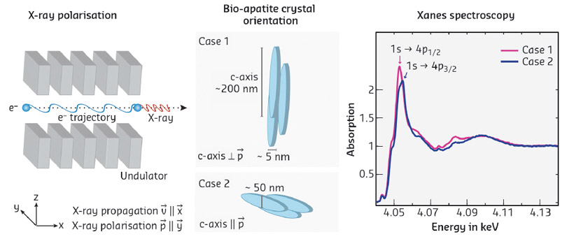
Fig. 48: Schematic view of the impact of the crystal orientation with respect to the polarisation vector on the spectral shape. The X-ray beam is generated by undulators resulting in a polarisation vector parallel to the horizontal plane. In bone apatite, crystals are arranged with the rotation axis perpendicular to the central blood vessel canals. The modulating crystal c-axis angles with respect to the polarisation vector result in a different spectral shape of the corresponding absorption spectra. |
Spectral differences, well pronounced in the XANES white line (related to 1s to 4p transitions of Ca), were observed in FF XANES (Figure 48). Using mathematical simulations and site-matched polarised Raman spectroscopy, which has been shown to be sensitive to the orientation of bone components, spectral differences could be associated with angular absorbance dependency of the Ca containing mineral crystals (Figure 49).
 |
|
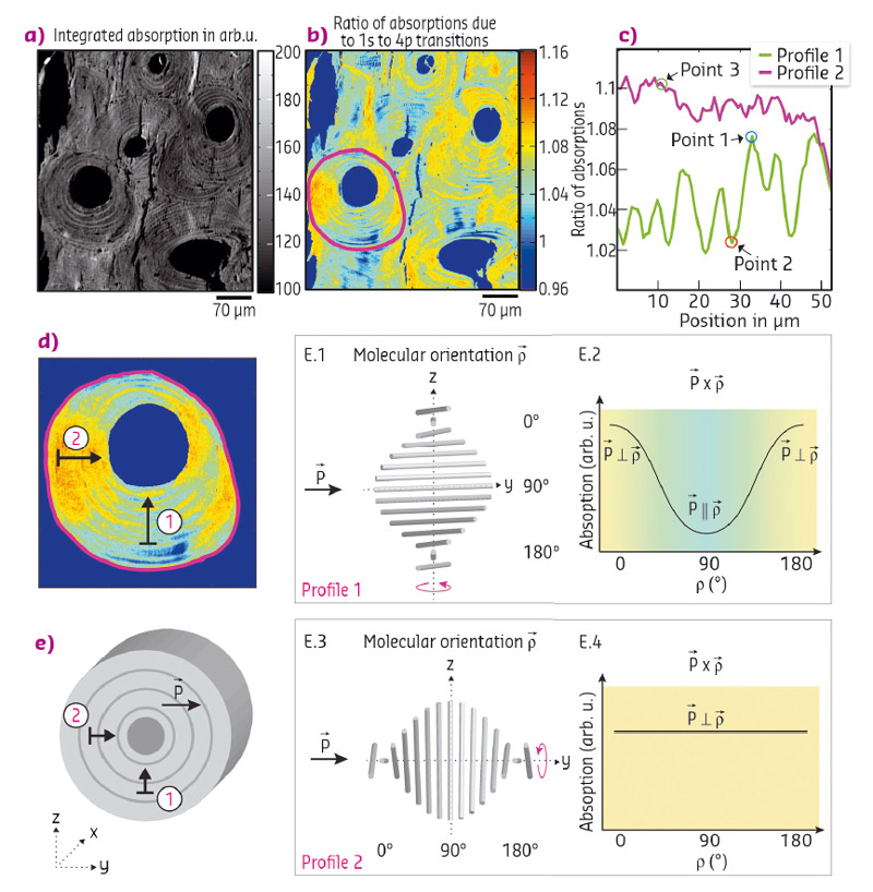
Fig. 49: FF-XANES investigation of bone structural unit section. Pixel size is 0.7×0.7 µm2. a) Integrated absorption of the full XANES energy range and b) Ratios of the absorption amplitudes corresponding to the 1s to 4p transitions. c) Line plots along a vertical and horizontal direction (as indicated in (d) reveal the orientation dependence of the lamellar pattern. d) Enlargement of a sub-region as indicated in (b). The origin of the modulations is outlined in (e). Comparison of simulated model and experimental data: The X-ray beam is polarised in the x-y plane and propagating in x- direction. The cross-product between polarisation vector and mineral orientation for a twisted plywood arrangement is shown in E.2. As the cross-product is not constant, it potentially explains the variations seen in profile 1 shown in (c). In contrast, the polarisation vector is perpendicular to all molecular orientations within ROI2, therefore the cross-product is unvarying for all mineral orientations (E.4) explaining the far less pronounced contrast in profile 2 shown in (c). |
Our data suggested that neither the anatomical site nor the pathology affected the averaged spectral shape of the XANES spectra. Instead, we could demonstrate that the spectral variances were dominated by angular dependent absorption effects. It is indeed crucial to differentiate between structurally and chemically induced spectral alterations. The fine structure modulations observed are of high interest for structural analysis of mineral crystal orientation in highly-structured tissue matrices.
The combination of fast acquisition time, large field of view and high lateral resolution makes FF-XANES spectroscopy a potential alternative to standard X-ray diffraction techniques and diffraction-limited methods such as polarised Raman spectroscopy. Furthermore, collecting µXRF maps in scanning mode at only two distinct energies is already sufficient to derive information on crystal orientations. This configuration is of particular interest when XANES cannot be acquired in transmission mode. Finally, we anticipate that this approach will be valuable for the study of other highly-organised bio-materials, such as teeth, and that it could be used not only to map crystal orientation but also to evaluate alterations of apatite crystal states such as changes of the c-lattice parameters.
Principal publication and authors
Full-field calcium K-edge X-ray absorption near-edge structure spectroscopy on cortical bone at the micron-scale: polarization effects reveal mineral orientation, B. Hesse (a), M. Salome (a), H. Castillo (a), M. Cotte (a,b), B. Fayard (a), C. Sahle (a), W. De Nolf (a), J. Hradilova (c,d), A. Masic (e), B. Kanngießer (f), M. Bohner (g), P. Varga (h), K. Raum (c) and S. Schrof (c), Analytical Chemistry 88, 3826–3835 (2016); doi: 10.1021/acs.analchem.5b04898.
(a) ESRF
(b) Sorbonne Universités, UPMC Univ Paris 06, CNRS, UMR 8220, Laboratoire d’archéologie moléculaire et structurale (LAMS), Paris (France)
(c) Charité - Universitätsmedizin Berlin (Germany)
(d) Czech Technical University in Prague, Faculty of Nuclear Science and Physical Engineering (Czech Republic)
(e) MIT, Department of Civil and Environmental Engineering, Cambridge (USA)
(f) Technical University of Berlin, Institute for Optics and Atomic Physics (Germany)
(g) RMS Foundation, Bettlach (Switzerland)
(h) AO Research Institute Davos (Switzerland)
partners


European Synchrotron Radiation Facility - 71, avenue des Martyrs, CS 40220, 38043 Grenoble Cedex 9, France.