- Home
- Holographic diffraction imaging with hard X-rays
Holographic diffraction imaging with hard X-rays
Nanoscience needs the ability to characterise the structure of objects on the nanoscale. For several reasons, hard X-rays are very attractive for this purpose: a wavelength in the order of ≈ 1 Å allows excellent spatial resolution; the high penetration into matter permits the study of thick samples or even buried structures in an easy-to-realise experimental setup; and with the upcoming hard X-ray free-electron lasers, the achievable time resolution will reach the femtosecond (fs) regime. Here, we demonstrate how coherent hard X-rays can be used for a determination of the absolute electron density of a lithographically tailored gold nanostructure (the letter P) from a single diffraction experiment, yielding both the shape and the height of the sample. We combine Fourier transform holography [1], which – by a single Fourier transform – gives an unambiguous image of the sample structure, with iterative phase retrieval procedures [2] that enable us to push the spatial resolution toward the diffraction limit, which is determined by the maximum photon momentum transfer.
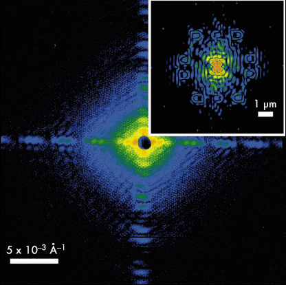
In Fourier transform holography, phase retrieval becomes particularly simple since, in a typical setup, the object and a spatially nearby reference are coherently illuminated. A single Fourier transform of the recorded hologram yields the convolution of the object and reference amplitudes, where the spatial resolution achievable is comparable to the reference source size [1]. In the present case 5 gold dots with about 175 nm diameter were placed on a circle of 2.5 µm radius around the gold nanostructure, each acting as reference source in the scattering process.
 |
|
Fig. 53: Recorded hologram of a gold nanostructure – the capital letter P – surrounded by 5 reference scatterers. The inset shows the central part of the modulus of the Fourier transform of the recorded diffraction intensities, imaging the object and its rotated copy 5 times each. Both images are shown on a logarithmic pseudocolour scale. |
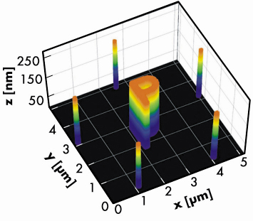
The corresponding hologram, which is shown in Figure 53, was recorded at beamline ID10C using coherent 8 keV photons. A single Fourier transform of the measured intensities gives the spatial autocorrelation of the overall object, including the cross-correlation between the object and the dots, which yields the object shape directly and its complex conjugate (a 180°-rotated copy) for each dot, as can be seen in the inset of Figure 53. The individual images are averaged to improve the statistics and the result – already comprising a qualitative image of the object – is fed into further iterative phase retrieval algorithms that are commonly used in coherent diffraction imaging [2]. Carrying out such phase retrieval runs yielded an image of the sample’s electron density projected along the beam direction with ≈ 25 nm resolution. Knowing that the sample under investigation was made of gold and exploiting the fact that the scattering process can be described as Thomson scattering, it is straight forward to derive a mean sample height, which would be the correct height if the sample were flat. Actually, in the present case we can tell how well the assumption of a flat sample topography is, since this information is encoded in the phases of the hologram’s Fourier transform. For our sample, a homogeneous height is confirmed with the actual value of ≈ 235 nm, as illustrated in Figure 54.
 |
|
Fig. 54: Visualisation of the object geometry (letter plus reference dots) as determined from the electron density profile. |
In conclusion, we have shown that hard X-ray holographic diffraction imaging is an excellent technique for determining electron density profiles on the nanoscale. A combination of the Fourier transform holography result and iterative phase retrieval methods made it possible to push the spatial resolution toward the diffraction limit. By virtue of this concept, we determined the absolute electron density and derived both shape and height of a lithographic gold nanostructure. This approach with coherent hard X-rays is ideally suited for applications in materials science, where samples can be thick, may consist of buried structures, or have to be measured under extreme conditions such as high pressure. Finally, we envision experiments of similar type to be carried out at future hard X-ray free-electron laser sources, where diffraction patterns can be collected within a single shot at the time scale of ≈ 10 fs, opening fascinating possibilities for imaging fast dynamic processes.
Principal publication and authors
L.-M. Stadler (a), C. Gutt (a), T. Autenrieth (a), O. Leupold (a), S. Rehbein (b), Y. Chushkin (c), G. Grübel (a), Phys. Rev. Lett. 100, 245503 (2008).
(a) HASYLAB at DESY, Hamburg (Germany)
(b) BESSY, Berlin (Germany)
(c) ESRF
References
[1] S. Eisebitt, J. Lüning, W.F. Schlotter, M. Lörgen, O. Hellwig, W. Eberhardt, J. Stöhr, Nature (London) 432, 885 (2004).
[2] H.N. Chapman, A. Barty, S. Marchesini, A. Noy, S.P. Hau-Riege, C. Cui, M.R. Howells, R. Rosen, H. He, J.C.H. Spence et al., J. Opt. Soc. Am. A 23, 1179 (2006).
partners


European Synchrotron Radiation Facility - 71, avenue des Martyrs, CS 40220, 38043 Grenoble Cedex 9, France.