- Home
- Phase Contrast X-ray Microscopy at 4 keV Photon Energy with 60 nm Spatial Resolution
Phase Contrast X-ray Microscopy at 4 keV Photon Energy with 60 nm Spatial Resolution
Recent progress in nanotechnology makes it possible to fabricate high aspect ratio Fresnel zone plates with nearly theoretical diffraction efficiency. For the multi-keV photon energy range, such zone plates are made of gold and are used as condenser and objective lenses. They permit X-ray imaging with high spatial resolution and short exposure times. Similar to conventional light microscopy, the resolution of an X-ray microscope is best when illuminating the object with a condenser, whose aperture is matched to the aperture of the imaging objective. The full-field X-ray microscope installed at the ID21 beamline, operating at 4 keV photon energy with condenser and objective zone plates of matched apertures and 50 nm outermost zone width, currently achieves 60 nm spatial resolution. It was realised that many scientifically-relevant samples provide only weak absorption contrast in the multi-keV photon energy range, whereas much higher contrast could be achieved in the phase contrast mode of an X-ray microscope [1].
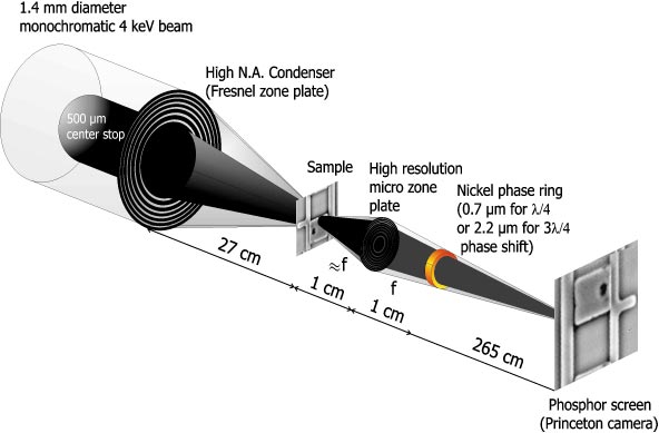
According to Zernike, phase contrast images are obtained in a full-field microscope by inserting a phase plate in the objective's back focal plane, which only shifts the undiffracted light's phase. In the novel Zernike-type phase contrast mode of the X-ray microscope, a ring-shaped condenser in combination with a central beam stop (see Figure 102) provides a hollow-cone illumination of the object. This also creates an image field free of zero order radiation from the zone plate objective and allows the introduction of a ring-shaped phase plate matched to the location of the undiffracted light, while light diffracted by sample structures remains nearly unaltered. Thereby, the phase information of the object is transformed into an intensity modulation in the image plane. In the Zernike-type phase contrast setup, even the image contrast of weakly phase-shifting samples can be tuned to almost any value by employing optimised phase rings, which also strongly attenuate the undiffracted light [2].
 |
|
Fig. 102: Schematic of the optical setup of the X-ray microscope (TXM) in Zernike phase contrast mode. A zone plate condenser focuses the monochromatised undulator beam onto the object and a micro zone plate objective forms a magnified image on a phosphor-coupled CCD camera. The phase ring selectively phase shifts the undiffracted light of the object. |
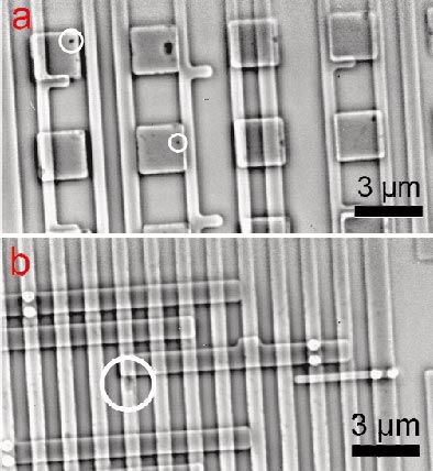
In the multi-keV photon energy range, phase-contrast X-ray microscopy is a new tool for investigations in biology, materials and environmental sciences. As an example for high-resolution phase contrast imaging in the Zernike mode, we apply this technique to integrated circuits. State-of-the-art microprocessors, which operate at frequencies in the GHz range, require the integration of more than 100 million transistors which have to be connected by metal lines buried in dielectrics. As the line width of the interconnects continue to shrink, the formation of voids in interconnects induced by high current densities (electromigration) during integrated circuit operation can cause an open circuit or an increase in resistance, resulting in malfunction or speed degradation. Phase-contrast X-ray micrographs of advanced microprocessor test structures with copper interconnects and vias are shown in Figure 103 and Figure 104. As demonstrated with these images, phase-contrast X-ray microscopy is a powerful new technique for integrated circuit process parameter control and defect analysis.
 |
|
|
 |
|
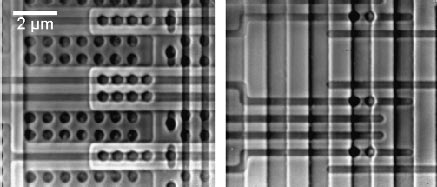
Fig. 104: Negative phase-contrast images of microprocessor structures from different regions on a chip, imaged with the ID21 transmission X-ray microscope. Note that the contrast is reversed compared to the X-ray micrographs taken in positive phase contrast shown in Figure 103(a) and (b). |
Future perspectives for this technique include the development of new phase-contrast microscopes operating at higher energies, driven by the possibility to study weakly-absorbing structures in thick samples with high spatial resolution. In addition, due to the large depth of field of X-ray objectives operating in the keV photon energy range, high-resolution tomography based on X-ray microscope images is possible.
The authors are indebted to E. Anderson and B. Harteneck from the Center for X-ray Optics, LBNL, Berkeley, for fabricating the phase rings.
References
[1] U. Neuhäusler, G. Schneider, W. Ludwig, D. Hambach, Proceedings of the 7th International Conference on X-ray Microscopy, Journal de Physique IV, in press
[2] G. Schneider, U. Neuhäusler, W. Ludwig, D. Hambach, submitted
Principal Publication and Authors
U. Neuhäusler (a), G. Schneider (b), W. Ludwig (a,c), M.A. Meyer (d), E. Zschech (d), D. Hambach (e), J. Phys. D, in press.
(a) ESRF
(b) Center for X-ray Optics, Lawrence Berkeley National Laboratory (LBNL), Berkeley (USA)
(c) now with INSA de Lyon, Villeurbanne (France)
(d) AMD Saxony LLC & Co. KG, Dresden (Germany)
(e) Institut für Röntgenphysik, Universität Göttingen (Germany), now with EPO, Munich (Germany)
partners


European Synchrotron Radiation Facility - 71, avenue des Martyrs, CS 40220, 38043 Grenoble Cedex 9, France.