- Home
- Synchrotron Microtomography of Fatigue Crack Closure
Synchrotron Microtomography of Fatigue Crack Closure
The incidence of fracture surface contact in the wake of fatigue cracks, and the associated attenuation of cyclic displacements, is known to be critical to crack propagation resistance. Whilst such surface contact (or 'closure') has been widely investigated over the last 20-30 years, measurement methods remain the subject of ongoing controversy, with the most widely established methods (compliance based) offering little micromechanical insight, or indeed any direct information on the active crack tip region. Synchrotron microtomography has been identified as a uniquely powerful method to obtain in situ understanding of plane strain crack tip behaviour [1]. Resolution levels have however been limited to date (~ 6 µm), requiring sub-voxel interpolation to be used in the crack tip region. Recent work has demonstrated the ability of high-resolution synchrotron microtomography to image the fine details of fatigue cracks together with surrounding microstructural features in Al-alloys with a 0.7 µm resolution [2]. This resolution has been used in the present work to image crack closure in a 2024 alloy, carried out on beamline ID19. Work has been particularly carried out using the damage tolerant airframe aluminium alloy, AA2024-T351. By extracting relatively small tomography samples from the crack tip of conventional fatigue test coupons, results representative of aerospace engineering performance have been obtained. In situ straining facilities have been used to assess the progressive loading and unloading process.
 |
|
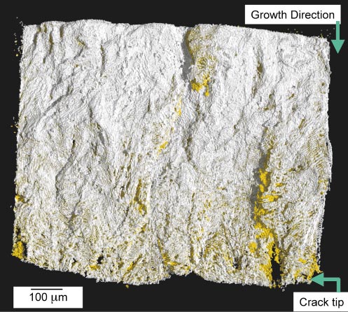
Fig. 91: Thresholded crack volumes obtained at maximum and minimum loads. |
Tomography results have been used for both direct imaging of crack paths and 3D image analysis to extract the crack volume. Figure 91 shows 3D crack volumes extracted in the loaded and unloaded conditions, rendered and viewed perpendicular to the crack plane. The white layer represents the unloaded crack, superimposed over the same crack at maximum load in yellow. As representations of the crack volume in each case, discontinuities in the surfaces represent either unbroken ligaments in the crack wake, or regions of fracture surface contact. The regions appearing in yellow clarify areas in the crack wake that have gone from a closed to open condition on loading. Such results identify distributed, relatively small points of contact close to the crack tip (of the order of 10 µm across), and large contact areas (of the order of 100 µm) occurring to greater distances behind the crack tip. The larger contact areas and their apparent association with ridges running parallel to crack growth direction is consistent with previous reports, whilst the distribution of small contact points approaching the crack tip has not been seen previously. Quantitative assessment of contact areas has confirmed the progressive nature of closure processes during crack loading and unloading. This highlights the intrinsic simplification and deficiency involved in the widespread use of a single valued closure point to quantify crack driving forces.
 |
|
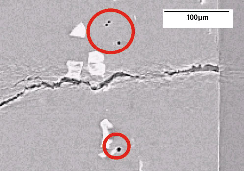
Fig. 92: Two-dimensional slice of a tomographic reconstruction showing crack and fine porosity used to quantify local deformations. |
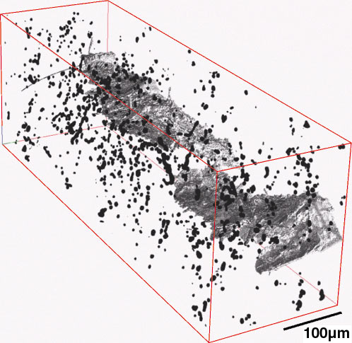
Given the available spatial resolution, a unique feature of the present work has been the quantitative use of micro-porosity and intermetallic particles associated with the underlying material for compliance/displacement measurements at a microstructural scale (Figure 92) [3]. Figure 93 highlights the three-dimensional distribution of such features around the crack tip, as have been used to measure local stress intensity factors around the crack tip, providing explicit information on changes in crack driving force as a function of location within the bulk sample.
 |
|
Fig. 93: Three-dimensional arrangement of particle and micro-void locations about the crack tip used for stress intensity calculations. |
References
[1] A. Guvenilir, T.M. Breunig, J.H. Kinney and S.R. Stock, Phil. Trans. R. Soc. Lond. A, 357, 2755-2775 (1999).
[2] W. Ludwig, J-Y. Buffière, S. Savelli and P. Cloetens, Acta. Materialia 51(3), 585-598 (2003).
[3] H. Toda, I. Sinclair, J-Y. Buffiere, E. Maire, T. Connolly, M. Joyce, K.H. Khor, and P.J. Gregson, Phil. Mag, submitted.
Authors
K.H. Khor (a), M. Joyce (a), T. Connolley (a), H. Toda (b), I. Sinclair (a), E. Maire (c), J-.Y. Buffiere (c).
(a) Southampton University (UK)
(b) Toyohashi University (Japan)
(c) INSA (France)
partners


European Synchrotron Radiation Facility - 71, avenue des Martyrs, CS 40220, 38043 Grenoble Cedex 9, France.