- Home
- Users & Science
- Scientific Documentation
- ESRF Highlights
- ESRF Highlights 2001
- Micro-analysis and Imaging
- New Results in X-ray Fluorescence Computed Microtomography
New Results in X-ray Fluorescence Computed Microtomography
XFCMT is a non-destructive, non-invasive imaging method that has started to play an increasing role in microanalysis during the last three years [1]. It complements transmission imaging by offering the much-needed elemental sensitivity down to trace element concentrations with the same micrometre-sized spatial resolution.
The method has been described extensively [1 and references therein], and currently ID22 performs fluorescence tomography with resolutions as high as 1 micrometre, using the microprobe setup with compound refractive lenses with fluxes as high as 109 to 1010 ph/s in the beamspot, for energy in the range 14 25 keV, while PINK beam mode allows flux increases of 10-20 times with the expected slight degradation in the bandwidth.
The sample imaged was a micro-fragment from the Tatahouine meteorite, fallen in 1931 and promptly stored at the Natural History Museum in Paris. The grain analysed was recently retrieved from the original site and compared to the pristine ones retrieved in 1931. A series of experiments in SEM and TEM [2] were performed, in order to study the remnants of pleomorphic bacteria present in fractures and fault lines of the meteorite.
The grain was placed in a sealed thin (![]() 10 µm thick) silica capillary to guarantee the non-destructiveness of the measurement since we wanted to perform IR spectroscopy afterwards. As the scattering peaks produced a non negligible background, it was necessary to use the AXIL fitting package for online data analysis, particularly for the low energy lines which are subject to self-absorption.
10 µm thick) silica capillary to guarantee the non-destructiveness of the measurement since we wanted to perform IR spectroscopy afterwards. As the scattering peaks produced a non negligible background, it was necessary to use the AXIL fitting package for online data analysis, particularly for the low energy lines which are subject to self-absorption.
The microbiological study of this meteorite revealed bacteriomorphs of sizes between 0.1 to 0.6 µm, which were obtained by cultures of the soil surrounding the grains. Hence, it was postulated that these bacteria appear at the fracture sites of the grain, following fluid circulation of carbonates from the soil due to terrestrial weathering. Therefore, our study aimed at identifying and locating non-invasively carbonate phases as well as pyroxenes and chromites specific to the grain. The grain was analysed through the silica walls of the capillary which posed no problems for the imaging of any elements with the exception of Si which was the main constituent of the capillary walls. In Figure 142 is shown the distribution of Fe, Cr and Ca particular to the phases expected, with a resolution of 2 µm using the ART reconstruction algorithm.
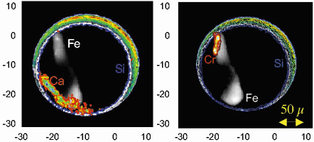 |
Fig. 142: Fluorescence tomograms of a micrometeorite grain inside a silica capillary. Reconstructions (using ART) of the K |
These analyses were completed by high-resolution (1 µm) 3D-transmission tomography that revealed the location and sizes of cracks and fractures as well as phases denser than the bulk of the grain. The tomographic set of data collected allowed us to obtain a rich image of the grain and to non-destructively characterise its morphology and structure, prior to the other analyses which require sample preparation and possibly alteration of the grain. This study is part of a CNES/NASA benchmark to establish the feasibility of such detailed analyses on Martian meteorites in the quarantine phase through the walls of a mini-P4 sample holder.
In the near future, the ID22 group will continue improvements of XFCMT, by upgrading both the experimental setup as well as the methodology of data collection, analysis and reconstruction, to enlarge the limits of the applicability of this technique.
References
[1] A. Simionovici, M. Chukalina, M. Drakopoulos, I. Snigireva, A. Snigirev, Ch. Schroer, B. Lengeler, K. Janssens and F. Adams, IEEE Trans. Nucl. Sci., 47, 2736 (2000).
[2] Ph. Gillet, J.A. Barrat, Th. Heulin, W. Achouak, M. Lesourd, F. Guyot and K. Benzerara, Earth & Planet. Sci. Lett. 175, 161 (2000).
Principal Publication and Authors
A. Simionovici (a), M. Chukalina (b), B. Vekemans (c), L. Lemelle (d), Ph. Gillet (d), Ch. Schroer (e), B. Lengeler (e), W. Schröder (f) and T. Jeffries (g), Developments in X-ray tomography III, ed. U. Bonse, SPIE 4503 (2001).
(a) ESRF
(b) IMT RAS, Chernogolovka (Russia)
(c) MITAC, Univ. of Antwerp (Belgium)
(d) École Normale Supérieure, Lyon (France)
(e) RWTH, Univ. of Aachen (Germany)
(f) IBI, FZ Jülich (Germany)
(g) British Natural History Museum, London (UK)
partners


European Synchrotron Radiation Facility - 71, avenue des Martyrs, CS 40220, 38043 Grenoble Cedex 9, France.