- Home
- Fast Strain Scanning with High Spatial Resolution
Fast Strain Scanning with High Spatial Resolution
Local strain/stress fields play a central role in the processing of polycrystalline bulk materials. They impact on the performance of components by superposition with load stresses, but also influence the microstructural development during processing. Gauge volumes down to micrometre dimension become achievable using microfocussed high-energy synchrotron radiation [1]. The thus accessible length scale includes gradients of grain averaged strain/stress fields of engineering interest in fine grained materials, e.g. in coatings or at buried interfaces, but also enables the isolation of individual grains and therefore the study of the underlying physical principles. Furthermore, in situ investigations become feasible by fast data acquisition.
The experimental technique consists of positioning a narrow slit behind the sample and observing the resulting diffraction pattern on a 2-D detector. The slit defines the spatial resolution parallel to the incoming beam and should be placed closely behind the sample. The scattering angles are measured by the 2-D detector. The strain is extracted from the central position of the diffraction peak. For the applications presented below, the slit was placed 10 cm behind the sample providing space for the sample environment. Due to the finite sample to slit distance, a coupling arises between the location of the gauge volume within the sample and the observed scattering angle. In an actual measurement, the sample is translated along the beam and images are taken at each sample position. Strains cannot be extracted directly from single images but the diffraction peak belonging to a certain gauge position must first be reconstructed from the recorded series of images.
The method was validated by measuring the in-plane strain profile across the surfaces of a shot-peened Al calibration sample. In this case, the same component could also be measured by the conventional crystal analyser technique. Figure 88 shows the good agreement of the measurements, the poorer resolution of the slit scan is due to the parallel orientation of the strain gradient to the elongated gauge volume.
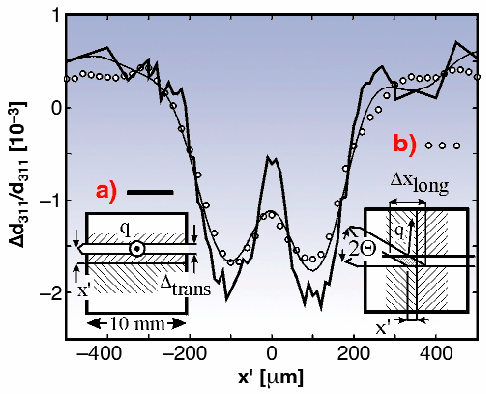 |
Fig. 88: In-plane strain profile of a test sample consisting of two shot-peened Al pieces. The sample geometry is shown in the insets.
|
Fast data acquisition has been achieved by the development of conical slit cells that enable the simultaneous observation of up to six complete diffraction rings. A biaxial strain state can be extracted from the distortion of the rings and the azimuthal intensity variation permits a texture analysis. Evaluation of the complete strain tensor and texture requires only sample rotation around one axis.
Torsion deformation is a complicated test case as the actual stress state is truly triaxial and depends strongly on the radial position within the sample. Figure 89 shows the sample geometry and the experimental setup. The strain profiles show a steep gradient at the sample centre and the presence of intergranular strains [2].
It was demonstrated that strain profiles can be measured by a slit imaging technique. In combination with a conical slit cell, extremely high data acquisition rates have been achieved which enable in situ profiling of the complete strain tensor, separation of macro- and intergranular strains, and texture determination. The above experimental work was performed at the ID11 beamline.
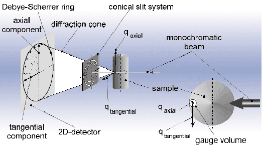 |
Fig. 89: 3-D strain mapping in a torsion sample using a conical slit cell.
|
References
[1] U. Lienert, H.F. Poulsen and Å. Kvick, AIAA Journal 39, (No. 4), (2001), accepted for publication.
[2] R.V. Martins, S. Grigull, U. Lienert, L. Margulies, and A. Pyzalla, Proc. of ICRS-6 Oxford (UK), 90-97 (July 2000).
Principal Publication and Authors
U. Lienert (a), R. Martins (a), S. Grigull (a), M. Pinkerton (b), H.F. Poulsen (c) and Å. Kvick (a), Mat. Res. Soc. Symp. Proc., 590, 241-246 (2000).
(a) ESRF
(b) Manchester Materials Science Centre (UK)
(c) Risø Nat. Lab., Roskilde, (Denmark)
partners


European Synchrotron Radiation Facility - 71, avenue des Martyrs, CS 40220, 38043 Grenoble Cedex 9, France.