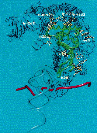
Figure 5
Fig. 5: Model of the protein S7-16S rRNA interaction. The S7 backbone derived from the crystal structure is colored green and the double-helical regions of the 16S rRNA in contact with the S7 are labelled. The highlighted sites are those of the Fe(II)-EDTA footprints. The anti-codon loop of the P site tRNA is coloured light blue and the model also includes a segment of the mRNA, coloured red, with its 5' end on the right hand side of the figure; the ribosome will move to the left along the mRNA as the translation proceeds.
partners


European Synchrotron Radiation Facility - 71, avenue des Martyrs, CS 40220, 38043 Grenoble Cedex 9, France.