- Home
- News
- General News
- Seeing the invisible:...
Seeing the invisible: ESRF Highlights 2006
14-02-2007
The ESRF welcomes more than 5400 scientists a year. In 2006, their experiments at the ESRF resulted in more than 1200 scientific publications that fill the pages of renowned journals. The ESRF Highlights is a unique document that brings together more than 70 accounts of the world-leading scientific research carried out at the facility by external users as well as in-house researchers.
Share
This year there are many Highlights from the new ESRF scientific areas of Environmental Sciences and Cultural Heritage. A broad range of synchrotron radiation techniques were employed for these studies. For example, X-ray microfluorescence was used to study the chemical composition of the pigments in Pompeian cinnabar paintings to help us understand the blackening of these beautiful treasures and microbeam synchrotron radiation diffraction was used to characterise the decay of ancient silk fabrics (Figure 1).
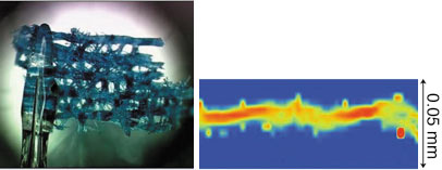 |
Fig. 1: Ancient silk fabric (left) and composite plot showing a single silk thread consisting of two twisted fibres (right). |
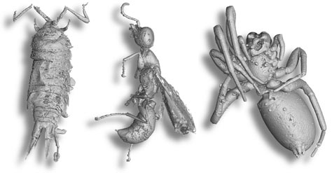
The phase or polarisation of the ESRF’s X-ray beam can be exploited to enhance the level of detail within a sample. Phase contrast X-ray synchrotron imaging was employed to look inside opaque amber resin, where a multitude of fossilised organisms could be found. Virtual 3D renderings of the organisms produced some surprisingly life-like creatures (Figure 2).
 |
|
Fig. 2: Examples of creatures preserved within the opaque amber. |
There are many Highlight articles that show the evolution of current techniques towards ever smaller sample sizes. These techniques take advantage of the small beam sizes that are obtained by focussing the beam to a micron or sub-micron spot size and analytical techniques that permit a synthesis of information from the data collected. The new MX microfocus beamline ID23-2 allows smaller crystal than ever before to be used for data collection. With X-ray standing wave imaging, the Surfaces and Interfaces group can even claim to be imaging on an atomic scale: the 3D reconstructed image of Mn doped GaAs shows the relative positions of the atoms (Figure 3).
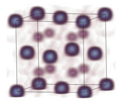 |
Fig. 3: Image of Mn within the GaAs unit cell. |
New spectroscopic methods are developed by the combination of techniques or with the sample under specific external conditions such as a high magnetic or microwave field. The article on X-ray Detected Magnetic Resonance presents a completely new technique to study materials within a microwave field that has great potential because of its element-selective nature. There is also a report on the first XRD results at beamline BM26B using a pulsed magnetic field up to 30T.
The examples given above represent only a few from the many exceptional accounts written by the ESRF's users and staff. Following on from these scientific highlights, the document then presents the report on the X-ray Source and the latest developments by the Machine Division. The Facts and Figures section includes a summary of the facility as well as a report on user operation.
To obtain a copy of the Highlights 2006 in printed format, please contact the Communication Unit. The on-line version is here: ESRF Highlights 2006.
partners


European Synchrotron Radiation Facility - 71, avenue des Martyrs, CS 40220, 38043 Grenoble Cedex 9, France.