- Home
- News
- General News
- “Double‑slit” experiment...
“Double‑slit” experiment on the nanoscale reveals hidden details of light‑matter interaction at the atomic level
14-04-2026
Scientists from the University of Göttingen and Hamburg and the ESRF have revealed details of light‑matter interaction at the atomic level using a ‘double‑slit’ experiment on the nanoscale in the first experiment using the nanoscope of ID14.
Share
Light is ‘refracted’ by transparent matter. We can admire this physical effect not only in rainbows but also in countless technical applications. Optical lenses, LCD screens, and broadband connections with fiber‑optic cables have become indispensable in everyday life. Ultimately, the entire modern information infrastructure relies on fast data exchange via optical fibres—and therefore on the refraction of light.
Refraction arises on the microscopic scale from the elastic interaction of light with individual atoms, whereby the light’s ‘oscillation phase’ is shifted slightly—a minute effect that, however, adds up over the enormous number of atoms. The strength of refraction—and thus the microscopic light‑atom interaction—can be measured very precisely with so‑called interferometers.
Designing interferometers for X‑ray radiation, or more precisely for ‘X‑ray light,’ is considerably more challenging. First, the effect is much weaker; second, the wavelength of X‑ray light is about 1 000 times shorter than that of visible light and even shorter than the typical inter‑atomic spacing in matter. This places extreme demands on the stability of an interferometer. At the same time, refraction is especially relevant for X‑rays because most matter appears transparent to them. This is exploited, for example, in X‑ray phase‑contrast imaging to acquire high‑resolution three‑dimensional images of biological specimens in a non-destructive way. Moreover, the interaction between X‑ray light and atoms is of particular interest: refraction provides information about the atoms contained in a sample and their immediate surroundings on a scale of a few nanometres.
 |
|
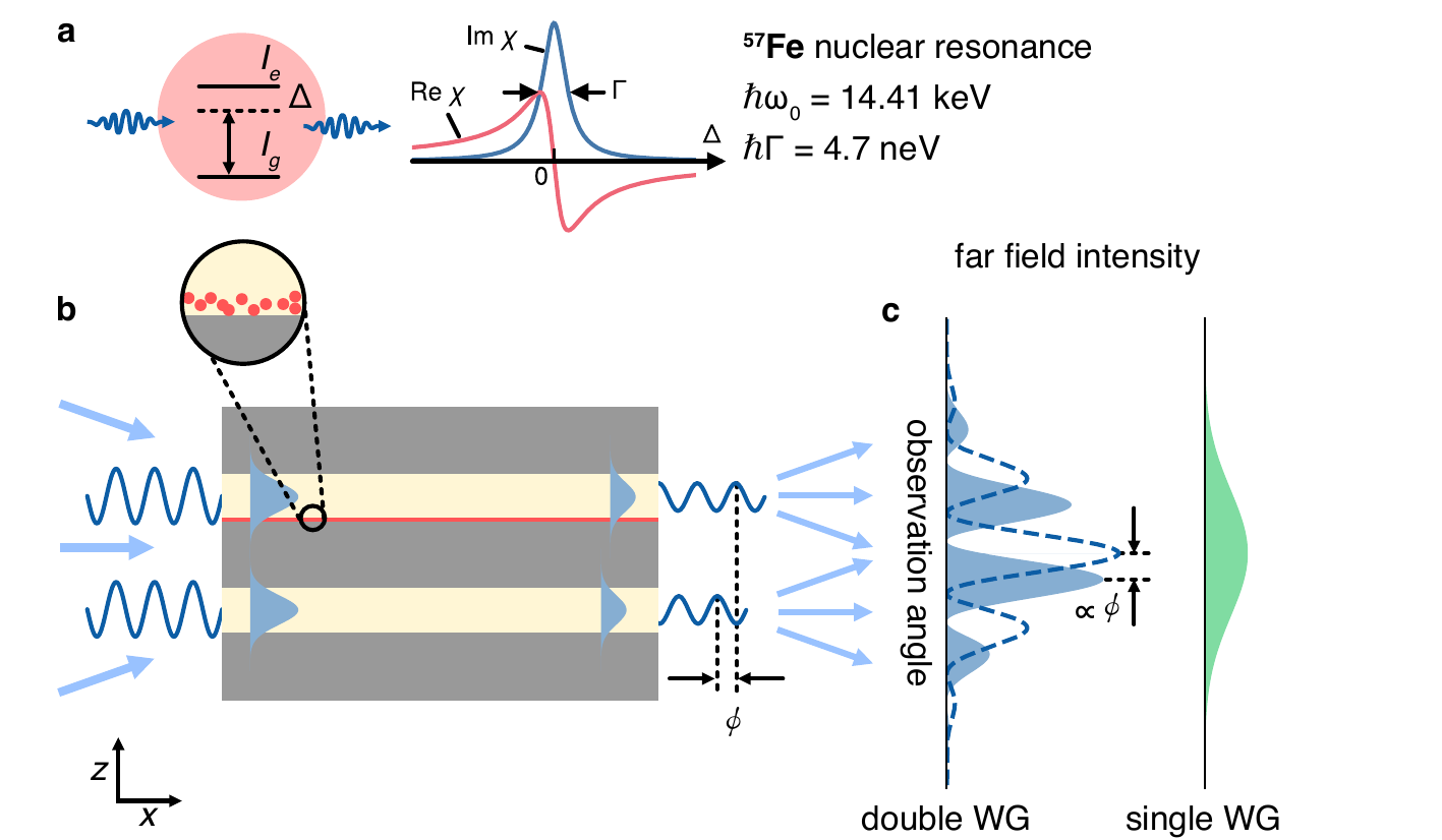
Principle of the interferometer. a) Interaction with the nuclei causes the light to be attenuated and its phase to be shifted. b) The phase shift becomes measurable through a double-slit interferometer, where one of the slits contains a thin layer of nuclei. c) The relative phase shift is converted to a lateral shift of the interference pattern in the far field. |
Researchers from Hamburg and Göttingen, together with partners in Grenoble, have, for the first time, demonstrated a miniaturised X‑ray interferometer that allows them to measure the interaction of X‑rays with atomic nuclei on the nanometre scale.
The interferometer’s principle is based on the famous ‘double‑slit’ experiment—the experiment that according to Nobel laureate Richard Feynman "has in it the heart of quantum mechanics". In this X‑ray interferometer the two slits are only 50 nm apart, roughly 1/1000 of the thickness of a human hair, making it probably the world’s smallest interferometer.
The experiments were carried out at the Nuclear Resonance beamline ID14 of the ESRF in Grenoble. In one of the two slits the atoms to be investigated were placed; in the present case an iron isotope (the Mössbauer isotope ⁵⁷Fe). “What is particularly fascinating is that the experiment was performed largely with single quanta of light ‘’or photons”, explains Leon Lhose, scientist at the University of Göttingen and corresponding author of the publication. Each photon traverses both slits simultaneously, interacting with the atomic nuclei in one of them and producing characteristic interference patterns behind the slits. From the measured patterns, the strength of the refraction at the atomic nuclei could be determined precisely, allowing conclusions about the light‑matter interaction.
“The experiment opens up rich perspectives as it shows how refraction provides additional information beyond simple attenuation in matter, especially when combined with atomic resonances”, says Lhose. It also lays the groundwork for systematic and precise determination of the refractive index of different elements for X‑ray radiation. In the future, ‘integrated optical circuits’ for X‑rays may even become conceivable.
Reference:
Lohse, L.M., et al. Nat. Photon. (2026). https://doi.org/10.1038/s41566-026-01892-5
Top image: Artistic rendering of the main data, the interference patterns. The distance from the viewer corresponds to the photon energy. At the nuclear resonance, a pronounced horizontal valley is clearly visible. In front and behind the resonance, the phase is shifted to opposing directions, which results in the shifted positions of the ridges.
partners


European Synchrotron Radiation Facility - 71, avenue des Martyrs, CS 40220, 38043 Grenoble Cedex 9, France.