- Home
- Users & Science
- Scientific Documentation
- ESRF Highlights
- ESRF Highlights 2017
- Enabling technologies
- X-ray imaging with random modulation: from the ESRF to laboratory sources
X-ray imaging with random modulation: from the ESRF to laboratory sources
By providing enhanced details of materials, phase contrast opened new perspectives for X-ray imaging at synchrotrons. To make this imaging modality accessible to a much broader community using compact laboratory sources, ESRF scientists have developed new methods to transfer phase imaging to low brightness sources.
Imaging is probably the most important application in the world of X-rays, considering its use as a non-invasive diagnostic in fields as different as medical imaging and security screening. Both 2D and 3D visualisation of samples are made possible using computed tomography due to the capacity of X-rays to penetrate matter. Very short wavelength is another property that makes X-ray imaging an invaluable probing tool to the scientific community. It permits much higher resolution than with visible light and with a greater depth of focus.
In this context, researchers are constantly looking for instrumentation and methods that could be implemented on moderate-cost X-ray laboratory equipment while simultaneously increasing the quantity and quality of information available. X-ray phase contrast imaging is a very attractive approach for this scientific case. Introduced a couple of decades ago, it showed sensitivities and signal-to-noise ratios much greater than for traditional absorption contrast employed in X-ray imaging since Röntgen’s discovery. However, although phase contrast is routinely obtained at synchrotrons, it is tedious to transpose onto laboratory systems due to the much lower brightness of these sources. For instance, propagation-based phase contrast imaging is easily observed at synchrotrons and does not require the implementation of any specific instrumentation. Unfortunately, such an approach is difficult to translate to laboratory sources due to the high divergence and low monochromaticity of these sources. In comparison, wavefront modulation-based techniques are better candidate methods to be transferred to these low-brightness sources. Speckle-based techniques, which fall into this category, are considered as a candidate of choice in this endeavour.
Work conducted at the ESRF test beamline BM05 over the last few years permitted the development of a multi-modal approach that offers the advantage of a very simple setup compatible with sources presenting short longitudinal and transverse coherence lengths. In a speckle-based method, the random modulation of the X-ray beam wavefront infers deflection by refraction in the sample. In contrast to the light experimental requirements of the approach, tracking the local displacement of the speckle modulation pattern using correlation algorithms calls for intensive numerical calculation. The random pattern, called speckle in the field of optics, is a modulation pattern very simple to obtain by using, for instance, a random mask made of grains, or, even more easily, using coherent light. In fact, when using a coherent synchrotron light source, speckle appears by interference effects as soon as an unwanted object or dust is present in the beam path, therefore it is often labeled as noise.
Several processing approaches based on the near-field speckle have been developed over the years. In the simplest speckle approach, one can achieve moderate sensitivity and resolution with a single exposure of a sample. In a scanning processing approach, several sample exposures achieve sub-micrometre resolution and nanoradian accuracy. Unfortunately, such performance was so far only possible at the cost of many sample exposures, especially in the case of highly divergent beams, hence preventing these methods spreading beyond the synchrotron community.
 |
|
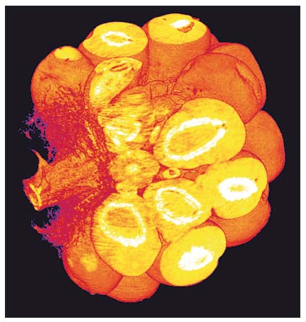
Fig. 150: Image reconstruction of a raspberry using the new speckle processing approach. |
With their new processing approach, scientists at the BM05 beamline found ways to perform the speckle-based imaging methods at laboratory sources. The new concept developed [1] consists of building, for each pixel, an oversampled basis of speckle pattern by collecting reference data prior to the sample measurement. When the sample is inserted in the beam, only very few exposures are made during a short scan. Then, the speckle pattern distorted by sample refraction is searched for its original position within the basis built earlier. Such processing greatly reduces the overall exposure time and overall data collection time without sacrificing either the resolution or the sensitivity of the method, even with highly diverging sources. Examples of data collected at the ESRF test beamline BM05 and processed with the new algorithm are illustrated in Figures 150 and 151.
 |
|
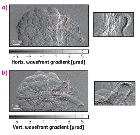
Fig. 151: a) horizontal and b) vertical wavefront gradient image of a raspberry obtained using the new speckle processing approach. The feature details in the insets show that the method used does not reduce the original resolution provided by the detector as can be the case with other wavefront modulation-based techniques. |
These developments are expected to promote the speckle-based approach at laboratory sources so that new fields of applications may benefit from the techniques of X-ray phase contrast imaging.
Authors
S. Berujon and E. Ziegler. ESRF
References
[1] S. Berujon and E. Ziegler, Physical Review A 95(6), 063822 (2017).
partners


European Synchrotron Radiation Facility - 71, avenue des Martyrs, CS 40220, 38043 Grenoble Cedex 9, France.