- Home
- Organic-inorganic X-ray detectors
Organic-inorganic X-ray detectors
Indirect flat-panel X-ray detectors provide limited resolution due to optical cross-talk. Here, a concept is presented whereby inorganic scintillating particles are embedded in an organic semiconductor matrix which effectively eliminates the optical cross-talk and increases the resolution. Experiments at the ESRF helped to gain knowledge on the device's morphology.
Radiography is an essential diagnostics branch in medicine. In projection radiography, the human body is exposed to X-rays and their transmitted intensity is captured by a detector. For decades radiologists have employed photographic paper as the detector. In spite of the introduction of solid state X-ray detectors, their full adoption has been hindered by the high cost. Researchers are now trying to develop low-cost and high-resolution flat-panel detectors for the energy range between 20 and 120 keV. Today's digital X-ray detectors mainly consist of a scintillator layer that converts X-rays into visible light and an imaging sensor that converts light into electrical signals. Apart from the high manufacturing costs, these detectors also have the disadvantage that the generated light propagates isotropically as it passes through the scintillator layer. As a consequence, the light hits a large number of adjacent pixels instead of hitting only one pixel of the image sensor. This results in a loss of spatial resolution.
 |
|
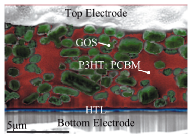
Fig. 70: Scanning electron microscopy (SEM) image of the device cross-section in false colour showing scintillating terbium-doped gadolinium oxysulfide (GOS: Tb) macroparticles (green) embedded in poly(3-hexylthiophene-2,5-diyl)) (P3HT) and phenyl-C61-butyric acid methyl ester (PCBM) bulk-heterojunction (red). HTL is the hole-transporting layer. |
In our hybrid approach, this problem has been solved by embedding the scintillator directly into a blend of organic semiconductors (bulk-heterojunction), which acts as a photodiode converting light into electrical charges (Figure 70). This gives the emitted light no opportunity to become widely spread and a high spatial resolution is obtained. Manufacturing costs are kept low thanks to effortless and effective spray deposition.
 |
|
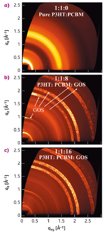
Fig. 71: GI-WAXS images of thermally annealed P3HT: PCBM: (GOS:Tb) films at different concentrations. |
Grazing-incidence wide-angle X-ray scattering (GI-WAXS) measurements performed at beamline BM28 (XMaS CRG) played an important role in understanding the device physics. Figure 71 shows diffraction images of P3HT: PCBM: GOS films at different concentrations. We observe a structural interplay between P3HT and GOS during thermal treatment. The amount of P3HT face-on lamellae increases with GOS concentration, which explains the increase in mobility measured by X-ray induced charges probed by X-ray charge carrier extraction by linearly increasing voltage (X-CELIV).
Principal publication and authors
X-ray imaging with scintillator-sensitized hybrid organic photodetectors, P. Büchele (a,b), M. Richter (c), S.F. Tedde (a), G.J. Matt (c), G.N. Ankah (d), R. Fischer (a,c), M. Biele (a,c), W. Metzger (a), S. Lilliu (e), O. Bikondoa (f,g), J.E. Macdonald (h), C.J. Brabec (c), T. Kraus (d), U, Lemmer (b) and O. Schmidt (a), Nature Photonics 9, 843–848 (2015); doi: 10.1038/nphoton.2015.216.
(a) Siemens Healthcare GmbH, Technology Center, Erlangen (Germany)
(b) Light Technology Institute and Institute of Microstructure Technology, Karlsruhe Institute of Technology (Germany)
(c) Institute of Materials for Electronics and Energy Technology (i-MEET), Friedrich-Alexander-University Erlangen-Nuremberg, Erlangen (Germany)
(d) INM – Leibniz Institute for New Materials, Saarbruecken (Germany)
(e) Masdar Institute of Science and Technology, Abu Dhabi (UAE)
(f) XMaS, The UK-CRG Beamline, ESRF, Grenoble (France)
(g) Department of Physics, University of Warwick, Coventry (UK)
(h) School of Physics and Astronomy, Cardiff University (UK)
partners


European Synchrotron Radiation Facility - 71, avenue des Martyrs, CS 40220, 38043 Grenoble Cedex 9, France.