- Home
- SAXS measurements explain the colour nanostructure relationship in feathers
SAXS measurements explain the colour nanostructure relationship in feathers
Structural colours are omnipresent in nature. Small-angle X-ray scattering has been used to understand the fine level of control that alters the size of the nanostructure in Jay feathers responsible for colour changes from blue to white. This control is made by regulating the duration of the β-keratin phase separation before vitrification.
The intensity and diversity of structural colours in nature is well-known. Only recently has the sophistication of the physics responsible for these optical effects been understood on the nanometre length scale [1,2]. Bird feathers have played an important role in our understanding of structural colour, as coloured feathers comprise nano-structured β-keratin materials that can be studied in great detail post mortem.
Two-dimensional Fourier analysis of electron microscopy images [3] showed that the blue colour in feathers is due to the constructive interference between light waves, coherently scattered by a nanostructured keratin–air matrix. Blue feathers function as Bragg reflectors. Scanning probe microscopy shows that the blue colour in the Jay’s feather is caused by a foam like structure.
 |
|
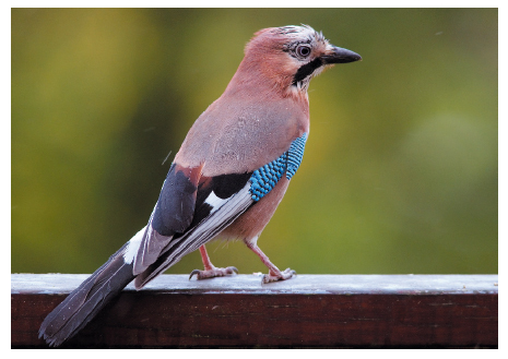
Fig. 96: Photograph of the Jay Garrulus glandarius showing the wing feathers with the periodic white blue black pattern (Open access image, credit: Luc Viatour/www.Lucnix.be). |
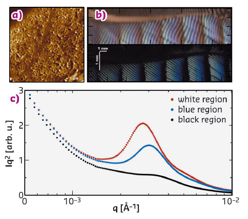
To probe the lengthscales in the blue and white pattern in the feathers of the Jay (Garrulus glandarius, Figure 96), we used the small-angle X-ray scattering (SAXS) setup at beamline ID02. The spatial colour map of the feather in Figure 97b was obtained by the Fourier transform of the SAXS data, which probes the periodic structure, which in turn determines the optical reflectivity. The data shows the periodic modulation in the size of the domain and consequently the colour as a function of position, which has not been seen in these nanostructures before. The colour in the top panel of Figure 97b was extracted from the SAXS data; it faithfully matches the colour in the optical image (lower panel) showing the link between nanostructure and optical reflectivity.
 |
|
Fig. 97: a) Scanning-probe image of the dark blue region showing the sponge-like morphology responsible for the optical properties. b) The colour derived from the X-ray determined structure (top panel) and the image of the area mapped (bottom panel) using X-ray scattering, showing the correlation of the nanostructure to the reflected colour. c) Representative Lorentz-corrected SAXS data for the different structural colour regions of a Jay feather barb (measured at beamline ID02). |
Given the q-range available in the SAXS setup, we were able to follow the large dynamic range in the structure that the feather barbs span. We used one dimensional correlation analysis to extract the real space morphology and transitions in size. This approach has also been used extensively in semi-crystalline polymers, using the CORFUNC software to examine the amorphous crystalline lamellae. With the only assumption that the sample is a two-phase system with differences in electron density, the auto-correlation function allows the period and the domain width of the nanostructure to be determined.
The optical structure responsible for the colour of Jay feathers is produced by a phase-separation process that is arrested at a late stage and the colour differentiation is achieved by controlling the duration of the phase-separation. This mechanism is likely to be widely shared amongst birds, reptiles and amphibians.
Principal publication and authors
Spatially modulated structural colour in bird feathers, A.J. Parnell (a), A.L. Washington(a,b), O.O. Mykhaylyk (c), C.J. Hill (d), A. Bianco (b), S.L. Burg (a), A.J.C. Dennison (e,f), M. Snape (a), A.J. Cadby (a), A.J. Smith (g), S. Prevost (h), D.M. Whittaker (a), R.A.L. Jones (a), A.R. Parker (i) and J.P.A. Fairclough (b), Nature Scientific Reports, 18317 (2015); doi: 10.1038/srep18317.
(a) Department of Physics and Astronomy, The University of Sheffield (UK)
(b) Department of Mechanical Engineering, The University of Sheffield (UK)
(c) Department of Chemistry, The University of Sheffield (UK)
(d) Department of Molecular Biology and Biotechnology, The University of Sheffield (UK)
(e) University Grenoble-Alpes, IBS, Grenoble (France)
(f) Institut Laue-Langevin, Grenoble (France)
(g) Diamond Light Source, Didcot (UK)
(h) ESRF
(i) Department of Zoology, Natural History Museum, London (UK)
References
[1] A.R. Parker, J. Opt. A: Pure Appl. Opt. 2, R15–R28 (2000).
[2] P. Vukusic et al., Science 315, 348–348 (2007).
[3] R.O. Prum et al., Nature 396, 28–29 (1998).
partners


European Synchrotron Radiation Facility - 71, avenue des Martyrs, CS 40220, 38043 Grenoble Cedex 9, France.