- Home
- Van Gogh’s Sunflowers: evidence of degradation of the chrome yellows
Van Gogh’s Sunflowers: evidence of degradation of the chrome yellows
The darkening of the yellow colours in Van Gogh’s Sunflowers (Van Gogh Museum, Amsterdam) has been strongly debated over the past few decades. This study presents firm evidence of the chemical alteration of chrome yellows in this iconic painting, outlining the regions with the highest risk of colour change due to the instability of the pigment used.
The fourth version of Sunflowers, painted by Vincent van Gogh in Arles in 1888-1889, comprises three similar paintings that are now on display at the National Gallery in London, the Seji Togo Memorial Sompo Japan Nipponkoa Museum of Art (Tokyo) and the Van Gogh Museum (Amsterdam), respectively. The vibrancy of these artworks was made possible by the use of chrome yellows (CYs), an early 19th century pigment class of which the hues ranged from lemon-yellow to orange-yellow. Despite their strong similarities, the three paintings feature different yellow tonalities with subtle variations in contrast among the sunflower petals, the background and the table area.
The extent to which different yellow shades are ascribable to Van Gogh’s stylistic choices (via the use of paint mixtures) and/or to unintended effects of colour changes (due to the degradation of original materials) has been questioned.
Earlier Cr K-edge μ-XRF/μ-XANES investigations at ID21 proved that the darkening of different CY types (PbCr1–xSxO4, with 0 ≤ x ≤ 0.8) in artificially aged paint models and microsamples taken from several Van Gogh paintings is due to a photo-redox process [1-4]. The formation of reduced Cr is more favoured for sulfate-rich, lemon-yellow, orthorhombic PbCr1–xSxO4 varieties of the pigment (with x > 0.4) than for the orange-yellow, monoclinic PbCrO4 that is the most lightfast of these materials [1,2].
In this study, non-invasive in situ spectroscopic investigations of the painting combined with synchrotron-based X-ray analysis of two microsamples were performed to assess the extent to which the Sunflowers version of the Van Gogh Museum (Figure 20a) contains lightfast PbCrO4 (denoted as LF-CY) and light sensitive S-rich PbCr1–xSxO4 (x > 0.4) (LS-CY), as well as to determine whether or not these pigments have been subject to a reduction process.
 |
|
Fig. 20: (a) Photograph of Van Gogh’s Sunflowers (Van Gogh Museum, Amsterdam) and (b) corresponding analysed micro-samples. (c) RGB composite MA-XRF maps of Pb/Cr/Zn. (d) Raman distribution of different CY types [LS-CY: light sensitive chrome yellow-PbCr1–xSxO4 (x~0.5); LF-CY: lightfast chrome yellow-PbCrO4; CO: chrome orange-(1-x)PbCrO4∙xPbO)]. “V” and “RL” indicate spots containing also vermilion and red lead, while the white circle shows the location where only red lead was identified. |
Non-invasive MA-XRF scanning combined with vibrational spectroscopic analysis (Figure 20c-d) revealed the use of different CYs, including the LF-CY and the LS-CY (x~0.5). Since these compounds were found in mixtures with other pigments (e.g. vermilion, zinc white, emerald green, red lead) as well as in unmixed form, the visual assessment of any colour change due to photo-reduction of the chromate is not straightforward.
The degradation state of the CY paint was evaluated by determining the Cr-oxidation state via Cr K-edge µ-XANES/µ-XRF analysis of two microsamples (Figure 20b) at ID21.
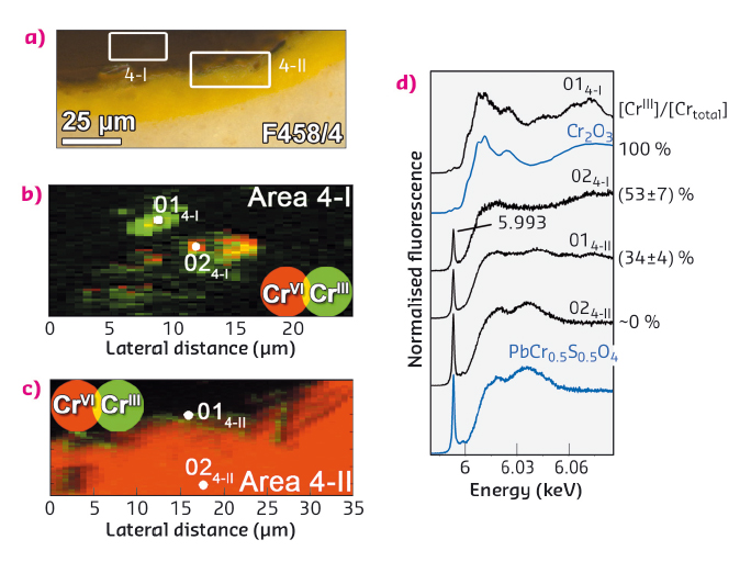
Clear indications of the presence of reduced Cr were found inside the varnish layer and at the varnish/paint interface of both these materials. At one location of sample F458/4 (from the light-yellow table; Figure 21a-b), a Cr-rich particle is completely reduced to the CrIII-state and the corresponding µ-XANES spectrum (Figure 21d: pt 014–I) resembles that of Cr2O3. In the area surrounding this particle, the relative abundance of CrIII was around 50% (pt 024–I). In another region (Figure 21c), about 35% of CrIII-species are present as a 2-3 µm thick layer right at the varnish/paint interface, while only CrVI-compounds constitute the yellow paint underneath (Figure 21d: pts 014–II-024–II). This pattern is very similar to that observed earlier in photochemically aged LS-CY (x > 0.4) paint models [2], suggesting that a colour change arising from CY reduction has occurred.
 |
|
Fig. 21: (a) Photomicrograph detail of sample F458/4 and corresponding (b-c) RG composite CrVI/CrIII chemical state maps. (d) µ-XANES spectra collected from areas indicated in (b-c). |
In conclusion, the results provide evidence of the chemical alteration of CYs in Van Gogh’s Sunflowers. Selected locations of the painting with the highest probability of colour change because of CrVI-reduction were identified, thus calling for careful monitoring in the future. The findings of this study, supported by those of our earlier work [1,2], open up the way to the elaboration of safer strategies for the light exposure of the painting.
Principal publication and authors
Evidence for degradation of the chrome yellows in Van Gogh’s Sunflowers: a study using noninvasive in situ methods and synchrotron-radiation-based X-ray techniques, L. Monico (a, b), K. Janssens (b), E. Hendriks (c), F. Vanmeert (b), G. Van der Snickt (b), M. Cotte (d), G. Falkenberg (e), B. G. Brunetti (a) and C. Miliani (a), Angewandte Chemie International Edition 127, 14129-14133 (2015) doi: 10.1002/anie.201505840.
(a) CNR-ISTM and Perugia University (Italy)
(b) Antwerp University (Belgium)
(c) Van Gogh Museum, Amsterdam (The Netherlands)
(d) ESRF
(e) DESY (Germany)
References
[1] L. Monico et al., Analytical Chemistry 85, 860-867 (2013).
[2] L. Monico et al., Journal of Analytical Atomic Spectrometry 30, 1500-1510 (2015).
[3] L. Monico et al., Analytical Chemistry 83, 1224-1231 (2011).
[4] L. Monico et al., Analytical Chemistry 86, 10804-10811 (2014).
partners


European Synchrotron Radiation Facility - 71, avenue des Martyrs, CS 40220, 38043 Grenoble Cedex 9, France.