- Home
- 6-dimensional diffraction contrast tomography and indexing by blind reconstruction
6-dimensional diffraction contrast tomography and indexing by blind reconstruction
Structural materials like metals commonly exhibit a crystalline microstructure, characterised by the presence of domains with distinct crystallographic orientation called grains. Depending on the thermo-mechanical process history of the material, these grains may be further divided into sub-structures created by the arrangement of lattice defects and giving rise to measurable distortion of the crystalline lattice. Diffraction contrast tomography (DCT) is an X-ray diffraction imaging technique that enables the non-destructive three-dimensional analysis of undeformed polycrystalline microstructures. A recently developed six-dimensional framework extended the applicability of DCT to the study of moderately deformed materials exhibiting intragranular misorientation of several degrees. Thanks to its fast acquisition protocoll, the technique enables time-lapse observations of processes like plastic deformation and grain coarsening in metallic or ceramic samples.
During acquisition of a diffraction contrast tomography (DCT) scan, a poly-crystalline sample mounted on a rotation stage is illuminated with a beam of monochromatic X-rays. Upon rotation, the various crystalline regions (grains) in the sample will repeatedly reach diffraction condition, and give rise to diffraction spots which are recorded on the detector positioned downstream of the sample. These diffraction spots, which represent geometrical projections of the grain volumes, will then be isolated (segmented), and used to identify and reconstruct the originating grains [1].
Due to the assumption of absent or negligible local misorientation, DCT has traditionally only been used for the structure determination of undeformed materials. However, many interesting problems in material science are linked to deformed microstructures. When materials present a non-negligible level of deformation, the diffraction spots are distorted, and tend to stay longer on the detector, transforming into diffraction blob volumes. Both their distorted nature, and the higher tendency to overlap, quickly degrade the quality of the traditional DCT reconstruction with increasing levels of grain deformation.
A new six-dimensional framework has extended the domain of applicability of DCT to include materials with moderate intragranular orientation spread by enabling the combined reconstruction of the shape and the local orientation inside each single grain. The six-dimensional nature of the method lies in the addition of three more degrees of freedom describing the local orientation for each spatial point and the simultaneous sampling of both position-space and orientation-space. The reconstruction is then formulated as a minimisation problem where additional prior terms that model the physical properties of the grains are used to cope with the increased complexity of the six-dimensional space.
In a more recent work [2], it was shown that the newly introduced framework is not only able to perform single grain reconstructions but it also enables the reconstruction of wider regions with similar orientation inside the analysed samples, along with the possibility to use raw images for the reconstruction of those regions. This means that previously difficult conditions will now be more approachable by DCT. Some examples include highly textured or moderately deformed materials where the diffraction spots have a much higher tendency to overlap and suffer from higher levels of distortion.
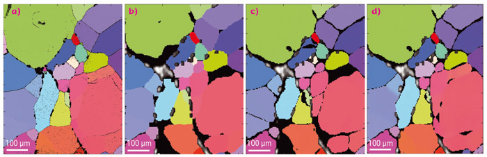
Figure 151a shows the surface a NaCl sample as observed by electron backscatter diffraction (EBSD), representing the surface crystalline structure, and its comparison with different approaches by DCT. Figure 151b is a dilated volume of a traditional 3D-DCT reconstruction and Figure 151c is an undilated 6D-DCT reconstruction.
 |
|
Fig. 151: Comparison of the reconstructions obtained with different approaches for the surface of a polycrystalline sample of rock salt (NaCl): (a) EBSD, (b) traditional DCT with a 2 voxels dilation, (c) 6D-DCT, (d) 6D-DCT with cluster reconstruction. |
Figure 151b and 151c are missing a few of the sub-grains that were successfully identified in the EBSD measurement. Figure 151d shows an extension of the 6D-DCT reconstruction where the adjacent grains with similar orientations and belonging to a cluster of grains have been reconstructed together in a larger region of the orientation-space. Including wider regions with similar orientations in the same reconstruction has enabled the reconstruction and identification of the regions that could not be indexed by a traditional DCT analysis.
More powerful computers would permit the analysis of even more deformed materials using DCT. Materials that exhibit higher degrees of overlap and distortion of the diffraction spots, could be analysed by simply extending the sampling to the full position and orientation spaces, thereby skipping the steps of image segmentation and grain indexing, and reconstructing the full sample at the same time. These developments, together with the fast acquisition speed of DCT and the experimental compatibility with other techniques such as phase contrast tomography, open up the possibility to perform time-lapse observations of plastic deformation, coarsening, phase transformation and crack propagation in polycrystalline structural materials.
Principal publication and authors
Reconstruction of local orientation in grains using a discrete representation of orientation space, N. Viganò (a,b,c), W. Ludwig (a,b) and K.J. Batenburg (d,c,e), J. Appl. Cryst. 47, 1826–1840 (2014); doi: 10.1107/S1600576714020147.
(a) MATEIS, INSA Lyon (France)
(b) ESRF
(c) iMinds-Vision Lab, University of Antwerp (Belgium)
(d) CWI, Amsterdam (The Netherlands)
(e) Universiteit Leiden (The Netherland)
References
[1] P. Reischig et al., J. Appl. Crystallogr. 46, 297 (2013).
[2] N. Viganò et al., Sci. Rep. accepted (2015).
partners


European Synchrotron Radiation Facility - 71, avenue des Martyrs, CS 40220, 38043 Grenoble Cedex 9, France.