- Home
- Microradian X-ray Diffraction in Colloidal Photonic Crystals and Liquid Crystals
Microradian X-ray Diffraction in Colloidal Photonic Crystals and Liquid Crystals
Self-assembly of colloidal particles into various crystalline and liquid-crystalline structures have been widely studied as a model of crystallisation of atomic and molecular systems as well as a route to photonic materials. The application of X-ray diffraction (XRD) to colloidal crystals is challenging due to many orders of magnitude difference between the size of a colloid and an X-ray wavelength. Angular resolutions of the order of a microradian are required to measure the intrinsic width of the diffraction peaks. Recent experiments performed at beamlines ID10A TROÏKA and BM26 DUBBLE met the challenge.
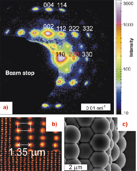
In particular, it is demonstrated that an angular resolution better than 2 µrad can be achieved with compound refractive X-ray optics. For instance, microradian diffraction can now be used to study crystals with spacings d larger than a micrometre. Such crystals can be used as templates to fabricate photonic materials for near-infrared telecommunication applications, which can manipulate light in a way similar to semiconductors manipulating electrons [1]. Figure 74 displays a diffraction pattern from a crystal of silica spheres (diameter 1.4 µm) with a body-centred tetragonal (bct) structure. The smallest diffraction angle of 2![]() 110 = 69 µrad corresponds to d110 = 1.35 µm. Despite the tiny diffraction angles, the pattern is clearly resolved and the width of the diffraction peaks can now be determined. Another advantage of these large-spacing crystals is that they can be studied not only by XRD, but also – after index matching with a suitable liquid – with 3D confocal microscopy, which is complementary to XRD. In addition, XRD can be applied to crystals that are strongly scattering or absorbing in the visible. This makes microradian XRD a unique tool to quantify stacking disorder, density of scattering centres, and the spatial extent of the positional order at different steps of the fabrication process. These results can now be used to improve the quality of the resulting photonic materials.
110 = 69 µrad corresponds to d110 = 1.35 µm. Despite the tiny diffraction angles, the pattern is clearly resolved and the width of the diffraction peaks can now be determined. Another advantage of these large-spacing crystals is that they can be studied not only by XRD, but also – after index matching with a suitable liquid – with 3D confocal microscopy, which is complementary to XRD. In addition, XRD can be applied to crystals that are strongly scattering or absorbing in the visible. This makes microradian XRD a unique tool to quantify stacking disorder, density of scattering centres, and the spatial extent of the positional order at different steps of the fabrication process. These results can now be used to improve the quality of the resulting photonic materials.
 |
|
Fig. 74: (a) Diffraction pattern from a colloidal bct crystal, measured at the ID10A branch of the TROÏKA beamline. The angular resolution is 5 µrad. (b) Projection of four hexagonal crystal layers obtained with 3D confocal microscopy from the original colloidal bct crystal (allows for quantitative comparison with XRD). (c) SEM image of the same crystal after drying and silicon infiltration (3D microscopy is no longer possible). |
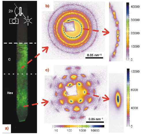
Moreover, correct identification of thermodynamic phases in colloidal suspensions crucially depends on the resolution [2]. Figure 75 presents results obtained in a concentrated suspension of colloidal gibbsite platelets (diameter 237 ± 49 nm, thickness 18 ± 3 nm), which form a columnar phase. The top part of the columnar phase consists of many small domains while a macroscopically large single-domain is observed in the bottom part, which looks well ordered at first sight. This is surprising since the particles have a very high diameter polydispersity. The nature of this surprising order is now seen clearly with the improved resolution. A radial peak broadening, which increases with the diffraction order, was detected. This indicates that the positional intercolumnar correlations are gradually lost. Such a fluid-like short-range positional order is typical for hexatic ordering [2], which is a compromise between the polydispersity-induced local frustrations and the long-range order. The significant difference in the spatial extent of the positional order, as well as a jump in the compressibility suggests that the single-domain is in fact a new liquid-crystalline phase: the hexatic columnar phase
 |
|
Fig. 75: (a) Photo of the sample in a Bragg-reflection geometry sketched in the inset. “C” and “Hex” denote hexagonal and hexatic columnar phases. Full XRD patterns and close ups of the (10) reflections in the multidomain hexagonal columnar (b) and the single domain hexatic columnar (c) regions, which are measured at BM26 DUBBLE. The angular resolution is 14 µrad. |
To summarise, small-angle X-ray diffraction from photonic colloidal crystals and liquid crystals has been performed with microradian angular resolution. This permits detailed characterisation of the structure and long-range order in strongly scattering photonic crystals with spacings larger than a micrometer [1]. We have also solved a long-standing problem of crystallisation in suspensions of highly polydisperse colloidal platelet-like particles [2].
References
[1] J.H.J. Thijssen, A.V. Petukhov, D.C. ’t Hart, A. Imhof, C.H.M. van der Werf, R.E.I. Schropp, A. van Blaaderen, to be published.
[2] A.V. Petukhov, D. van der Beek, R.P.A. Dullens, I.P. Dolbnya, G.J. Vroege, H.N.W. Lekkerkerker, Phys. Rev. Lett., 95, 077801 (2005).
Principal Publication and Authors
A.V. Petukhov (a), J.H.J. Thijssen (b), D.C. ’t Hart (b), A. Imhof (b), A. van Blaaderen (b), I.P. Dolbnya (c), A. Snigirev (d), A. Moussaïd (d), I. Snigireva (d), J. Appl. Cryst., in press (2006).
(a) van ’t Hoff laboratory, Utrecht University (The Netherlands)
(b) Soft Condensed Matter, Utrecht University (The Netherlands)
(c) BM-26 DUBBLE, ESRF; present address: Diamond Light Source (U.K.)
(d) ESRF
partners


European Synchrotron Radiation Facility - 71, avenue des Martyrs, CS 40220, 38043 Grenoble Cedex 9, France.