X-ray Imaging
Introduction
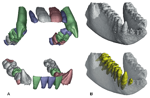
An increasing proportion of the experimental results obtained at the ESRF can be considered as "X-ray images". They are maps showing the variation of a physical quantity such as absorption, diffraction, fluorescence, photoemission, within the sample. Recent years have seen an enhancement of the possibilities of imaging/microanalysis techniques and their routine application to a range of topics including physical, medical, materials science and engineering, and also some new areas like geophysical, environmental, archaeological and biological studies. An example of the extension to new topics and scientific communities is given in Figure 132, which shows three-dimensional renderings of fossils from two Hominoid primates from Thailand. These fossils are of high paleontological significance as they are the closest-known relatives of the modern orangutan. The oldest is aged about 12 million years and the second about 7 million years. The comparison of the 3D reconstructions of the jaws of the two fossils allows us to conclude that the two species are very similar. For the second fossil, it was possible to perform a virtual 'extraction' of the teeth to study their roots (courtesy P. Tafforeau, J.-J. Jaeger and Y. Chaimanee [1,2]).
 |
|
Fig. 132: 3D rendering of two fossils from Thailand, relatives of the modern orangutan. a) virtual reconstruction of the jaws of the male of Khoratpithecus chiangmuanensis. Grey: real teeth. Pink: symmetrical equivalents of the real teeth. Blue: teeth from female after size correction. Green: extrapolated teeth. This species dates from about 12 million years ago. b) mandible of Khoratpithecus piriyai. This species dates from about 7 million years ago, but is very similar to the first one. In yellow: virtual extraction of the teeth showing their roots (Imaging was at ID19 and ID17 respectively, images courtesy P. Tafforeau, J.-J. Jaeger and Y. Chaimanee). |
The complementary microanalysis techniques already offered by the neighbouring "microscopy and microanalysis" beamlines ID21 (2-7 keV), ID22 (7-60 keV) and ID18F end station (6-28 keV) are being extended by the development of an infra-red microscopy end-station. Three examples show the present potential of these techniques: the characterisation of droplets and inclusions in "very old" ice from the Vostok lake, Antartica (First Direct Observation of Brine Droplets and Solid Inclusions in Accreted Ice from the Sub-glacial Lake Vostok (Antarctica)), the composition of 3500 million-year-old sea-water and its implications for the early stages of life (Modern Composition of 3,500 Million-year-old Seawater: Implications for Life on the Primitive Earth), and the variations of atmospheric sulphates recorded on stalagmites (Variations in Atmospheric Sulphate Recorded in Stalagmites).
Synchrotron radiation "wide field imaging", which includes radiography, microtomography, and diffraction topography, is increasingly used, the requested beam time being recurrently a factor 5 above the available time. Phase contrast imaging is one of the strong contributions of modern synchrotron radiation sources: the application chosen here shows its use for real-time investigations of the directional solidification of aluminium- based alloys (Application of Synchrotron X-ray Imaging to the Study of Directional Solidification of Aluminium-based Alloys). Microtomography is now implemented on a series of beamlines (BM05, ID15, ID17, ID19, ID22) to cover both academic, for example the study of liquid foams (X-ray Microtomographic Study of Liquid Foams), and industrial needs. One example of this last type of very applied research is for the study of the fermentation and baking of bread (In situ Study of the Fermentation and Baking of Bread Dough by X-ray Tomography). This subsection devoted to the study of materials is concluded by a in situ investigation of the roughness evolution during the growth of a Tungsten film (Roughness Conformity during Tungsten Film Growth: An in situ Synchrotron X-ray Scattering Study).
Synchrotron radiation based biomedical research is an important part of the X-ray Imaging Group's beamline activity, and more particularly of the dedicated beamline ID17. The quantitative comparison of various phase-contrast techniques to identify the most appropriate method for a given situation is the topic of the first contribution (Quantitative Comparison of Two Phase-contrast Techniques for Mammography). The parallel, and sometimes combined, development of diagnostic tools (e.g. using SAXS (Human Breast Tissue Characterisation with Small-angle X-ray Scattering), functional imaging and radiation therapy are active research fields. For instance, spectacular survival curves have been obtained for rats bearing glioma type brain tumours (Synchrotron Photoactivation of Platinum Against Brain Tumours). This is a highly encouraging result, which leads us to envisage its extension to clinical protocols. These programs are based on collaborations with local and European hospital teams, and the efficient use of the ESRF Biomedical Facility.
The techniques implemented at the ESRF also develop outside of the synchrotron radiation world. The common wish of the ESRF staff and of our users is to take full advantage of the specificities of third-generation synchrotron radiation facilities which are not available elsewhere. This implies continual development of the techniques, and lends particular importance to the section on advances in instrumentation.
The biomedical topics that are of fundamental or clinical relevance and for which synchrotron radiation techniques are the best tool are now well identified. The ESRF will concentrate on these topics, which will probably include clinical radiotherapy in the near future. To be able to answer the questions associated with modern science and techniques, synchrotron radiation microtomography is moving towards higher spatial and temporal resolutions. The temporal resolution (now in the 10 second range) is important for a wide range of time-evolving phenomena and should aim to reach the sub-second range. The spatial resolution, today in the 1 µm range, should be improved to the 50 nm scale ("nanoimaging" project), with the help of better focussing optics and detectors.
References
[1] Y. Chaimanee, D. Jolly, M. Benammi, P. Tafforeau, D. Duzer, I. Moussa and J.-J.Jaeger, Nature, 422, 61-65 (2003).
[2] Y. Chaimanee, V. Suteethorn, P. Jintasakul, B. Marandat and J.-J. Jaeger, Nature, 427, 439-441 (2004).
J. Baruchel
partners


European Synchrotron Radiation Facility - 71, avenue des Martyrs, CS 40220, 38043 Grenoble Cedex 9, France.