X-ray Imaging
Introduction
The X-ray imaging and microanalysis research effort, while of very wide application, exhibits a few easily-identifiable trends. These result from both the scientific demands and new technical capabilities such as improvements in the optics, detectors and computers and also in the stability of the X-ray source. These trends are the combination of medical imaging and therapy, the technical effort to produce higher spatial and temporal resolutions, and the implementation of adequate sample environments for in situ, real-time, experiments.
Biomedical research is an important activity, where there is an intimate link between X-ray imaging and radiotherapy. Our activity in this area is highlighted this year by the article on the treatment of brain tumours, an illness where the present number of treatments, and their efficiency, is unfortunately quite poor. The area of Microbeam Radiation Therapy (MRT) was described in last years Highlights. MRT is a very promising technique that is being actively developed at a preclinical stage. This development is carried out by identifying the best conditions to increase the survival, or even cure, the treated animals, and through more fundamental studies, at the cellular level, so that we can better understand the mechanisms of the action of the microbeams. Another radio-therapeutic approach is presented here: tomography-like radiotherapy combined with a contrast agent, which we call "Photoactivation Therapy", PAT. This type of radiation therapy uses a monochromatic beam with an energy optimised for an enhancement of the dose deposited at the level of the tumour; the choice of the energy depends on the heavy element (platinum or iodine) used as the dose-enhancing agent. The survival curves obtained are more than encouraging. A major effort is being made to gain simultaneously a deeper understanding of the biological mechanisms involved in PAT and to investigate its clinical impact and implementation. The second article in this chapter describes the application of the K-edge subtraction method, originally developed for coronary angiography, to functional lung imaging of an animal model, using Xe gas as contrast agent. This medical-imaging investigation allows a deeper understanding of the factors affecting ventilation, which are very important for a better treatment of diseases like asthma.
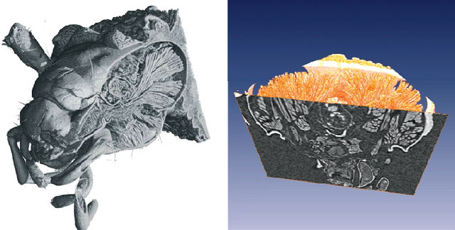
A second area of intense research is Imaging with very high spatial resolution. Figure 127 shows a recent example, where the inner part of an insect head has been visualised using tomography. The report on X-ray imaging of vascular networks, also emphasises the necessity of high spatial resolution: to understand the physiological role of the micro-vascularisation of the brain, visualisation of the vascular structure at the micrometre or even sub-micrometre scale is required. This was achieved for the first time using microtomography with an appropriate contrast agent.
 |
|
Fig. 127: Tomographic images showing the internal structures of the head of an earwig (Forficula auricularia), allowing the visualisation of the skeleton and muscles (Courtesy W.K. Lee, APS, O. Betz, Zoologisches Institut, Kiel and P. Cloetens, ESRF). |
As indicated above, an important feature of the X-ray imaging field is the very wide range of applications making use of the techniques. In addition to the biomedical applications, there are numerous applications from the fields of materials science and physics, which also benefit from the unique possibilities on the beamlines.
Porosities in quasicrystals are well visualised with phase-contrast imaging. The pore sizes were found to be much higher than those observed on crystals of the same composition. The evolution of the pores during a high vacuum annealing appears compatible with a vacancy diffusion mechanism; however, their origin remains a puzzle. This article also emphasises the importance of the sample environment, which allow in situ and real-time measurements.
Other highlight articles concern the analysis of grain compaction by X-ray microtomography this helps our understanding of the slow dynamics of non-equilibrium systems. Another example concerns the investigation of GaN, a very important material for new electronic devices. In this last case, the homogeneity is a key parameter, which was investigated in highly-doped Mn-GaN by using microanalytical techniques.
Synchrotron radiation microanalysis is an essential tool for many environmental studies, which, in addition to using the established techniques, involve development of new ones. Combined X-ray transmission, fluorescence and Compton scattering helical microtomography is a developing technique, which allows a precise quantitative reconstruction of the internal elemental composition and structure of the measured slice. This new method was applied to the investigation of a fly-ash particle. A new super-hard shock-induced polymorph of carbon was identified (probably resulting from a meteorite impact), that had not been predicted by any theoretical calculation. The degassing of sulphur during volcanic eruptions is a very important subject that remains unsatisfactorily explained. The evolution from Fe3+ to Fe2+ under conditions close to those in magmas during their ascent from the Earth's mantle to the surface was investigated by µXANES. Understanding the redox conditions furthers our knowledge of the whole process.
The intense activity in all of the areas mentioned will clearly lead to new developments in the near future. This is particularly true for radiation therapy and "nano-tomography". In this last case, an effort is foreseen to improve the spatial resolution to the 100 nm range, and in the longer term, to a few tens of nm.
J. Baruchel
partners


European Synchrotron Radiation Facility - 71, avenue des Martyrs, CS 40220, 38043 Grenoble Cedex 9, France.