Reviews
Reviews and perspectives on the use of X-ray microspectrosopy applied to environmental sciences
The ID21 Beamline has been a pioneer in the use of X-ray microspectrosopy applied to environmental sciences. The early experience of ID21 researchers and users on these materials gave them the oportunity to write their experiences and perspectives in several early articles, which paved the way for the use of synchrotron light in environmental and earth sciences. More recently, the use of synchrotron techniques on plant sciences, novel preparation methods like the use of cryo-techniques has been highlighted.
Soils
 |
J. Thieme, J. Sedlmair, S.-C. Gleber, J. Prietzel, J. Coates, K. Eusterhues, G. Abbt-Braun and M. Salomé, "X-ray spectromicroscopy in soil and environmental sciences", Journal of synchrotron radiation, 17, 149-157 (2010). |
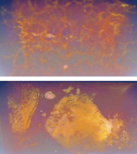
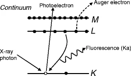
X-ray microscopy is capable of imaging particles in the nanometer size range directly with sub-micrometer spatial resolution and can be combined with high spectral resolution for spectromicroscopy studies. Two types of microscopes are common in X-ray microscopy: the transmission X-ray microscope and the scanning transmission X-ray microscope; their set-ups are explained in this paper. While the former takes high-resolution images from an object with exposure times of seconds or faster, the latter is very well suited as an analytical instrument for spectromicroscopy. The morphology of clusters or particles from soil and sediment samples has been visualized using a transmission X-ray microscope. Images are shown from a cryo-tomography experiment based on X-ray microscopy images to obtain information about the three-dimensional structure of clusters of humic substances. The analysis of a stack of images taken with a scanning transmission X-ray microscope to combine morphology and chemistry within a soil sample is shown. X-ray fluorescence is a method ideally applicable to the study of elemental distributions and binding states of elements even on a trace level using X-ray energies above 1 keV.
 |
E. Lombi and J. Susini, "Synchrotron-based techniques for plant and soil science: opportunities, challenges and future perspectives", Plant and Soil, 320, 1-35 (2009). |
Spectroscopic approaches to plant and soil sciences have provided important information for several decades. However, many of these approaches suffered from a number of limitations and drawbacks especially in terms of spatial resolution and requirements for sample preparation. The advent of dedicated synchrotron facilities, that allow the exploitation of the particular qualities of synchrotron radiation as a research tool, has revolutionised the way we approach the investigation of nutrients and contaminants in environmental samples. Various synchrotron-based techniques are currently available that permit such investigations in situ and at the molecular level. The continuous development of these techniques is delivering substantial gains in terms of sensitivity and spatial resolution which allows analyses of diluted samples at the sub-micron scale. This paper aims at providing an introduction to synchrotron radiation and to the fundamentals of some widely used synchrotron-based techniques, in particular X-ray absorption, fluorescence and tomography. Furthermore, examples are provided regarding the applications of synchrotron-based techniques in the field of plant, soil and rhizosphere research. Finally, current limitations and future perspectives of synchrotron techniques are discussed.
C. Schmidt, J. Thieme, U. Neuhäusler, C. Jacobsen, B. Kaulich, M. Salomé and J. Susini, "Spectromicroscopy of soil colloids", Journal de Physique IV, 104, 405-408 (2003).
A soil with a high organic content, a calcaric phaeozem, has been imaged in a hydrated state using the Stony Brook scanning transmission X-ray microscope at the NSLS, Brookhaven, USA. This experiment has been performed at the carbon K absorption edge to determine the distribution of organic matter within this soil. Spectromicroscopic methods using the known spectra of humics are helping to distinguish between different kinds of carbon in the sample. In addition, a marsh soil from Northern Germany has been analyzed at the sulfur K-absorption edge with the scanning X-ray microscope at the ESRF (ID 21), Grenoble, France. This soil contains a variety of sulfur containing components, which are attached to the soil colloids. A major source of these components is the microbial activity. In this soil it is possible to identify spectroscopically amino acids like methionin and cysteine and proteins, which are built up from those molecules, and inorganic components like sulfates and sulfides.
Plants
 |
H. A. Castillo-Michel, C. Larue, A. E. P. del Real, M. Cotte and G. Sarret, "Practical review on the use of synchrotron based micro-and nano-X-ray fluorescence mapping and X-ray absorption spectroscopy to investigate the interactions between plants and engineered nanomaterials", Plant Physiology and Biochemistry, 110, 13-32 (2017). |
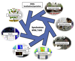
The increased use of engineered nanomaterials (ENMs) in commercial products and the continuous development of novel applications, is leading to increased intentional and unintentional release of ENMs into the environment with potential negative impacts. Particularly, the partition of nanoparticles (NPs) to waste water treatment plant (WWTP) sludge represents a potential threat to agricultural ecosystems where these biosolids are being applied as fertilizers. Moreover, several applications of ENMs in agriculture and soil remediation are suggested. Therefore, detailed risk assessment should be done to evaluate possible secondary negative impacts. The impact of ENMS on plants as central component of ecosystems and worldwide food supply is of primary relevance. Understanding the fate and physical and chemical modifications of NPs in plants and their possible transfer into food chains requires specialized analytical techniques. Due to the importance of both chemical and physical factors to consider for a better understanding of ENMs behavior in complex matrices, these materials can be considered a new type of analyte. An ideal technique should require minimal sample preparation, be non-destructive, and offer the best balance between sensitivity, chemical specificity, and spatial resolution. Synchrotron radiation (SR) techniques are particularly adapted to investigate localization and speciation of ENMs in plants. SR X-ray fluorescence mapping (SR-XFM) offers multi-elemental detection with lateral resolution down to the tens of nm, in combination with spatially resolved X-ray absorption spectroscopy (XAS) speciation. This review will focus on important methodological aspects regarding sample preparation, data acquisition and data analysis of SR-XFM/XAS to investigate interactions between plants and ENMs.
 |
C. Larue, H. Castillo-Michel, R. J. Stein, B. Fayard, E. Pouyet, J. Villanova, V. Magnin, A.-E. P. del Real, N. Trcera and S. Legros, "Innovative combination of spectroscopic techniques to reveal nanoparticle fate in a crop plant", Spectrochimica Acta Part B: Atomic Spectroscopy , (2016). |
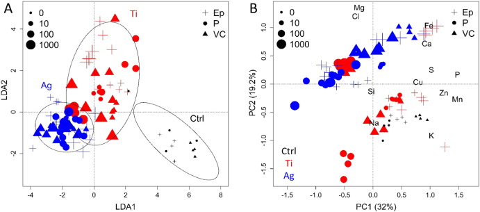
Nanotechnology is the new industrial revolution of our century. Its development leads to an increasing use of nanoparticles and thus to their dissemination. Their fate in the environment is of great concern and especially their possible transfer in trophic chains might be an issue for food safety. However, so far our knowledge on this topic has been restricted by the lack of appropriate techniques to characterize their behavior in complex matrices. Here, we present in detail the use of cutting-edge beam-based techniques for nanoparticle in situ localization, quantification and speciation in a crop plant species (Lactuca sativa). Lettuce seedlings have been exposed to TiO2 and Ag nanoparticles and analyzed by inductively coupled plasma spectrometry, micro-particle induced X-ray emission coupled to Rutherford backscattering spectroscopy on nuclear microprobe, micro-X-ray fluorescence spectroscopy and X-ray absorption near edge structure spectroscopy. The benefits and drawbacks of each technique are discussed, and the types of information that can be drawn, for example on the translocation to edible parts, change of speciation within the plant, detoxification mechanisms, or impact on the plant ionome, are highlighted. Such type of coupled approach would be an asset for nanoparticle risk assessment.
 |
G. Sarret, E. A. H. Pilon Smits, H. Castillo-Michel, M. P. Isaure, F. J. Zhao and R. Tappero, "Use of Synchrotron-Based Techniques to Elucidate Metal Uptake and Metabolism in Plants", Advances in Agronomy, 119, 1-82 (2013). |
Synchrotron techniques have become key components of the toolbox for studying the mechanisms involved in metal(loid) uptake and metabolism in plants. Most widely used techniques in this field include micro-X-ray fluorescence (μXRF) for imaging the distribution of elements in plant tissues and cells and quantifying them, and X-ray absorption spectroscopy (XAS) for determining their chemical forms. Recent advances in terms of spatial resolution, sensitivity and versatility of the sample environment have opened new perspectives for the study of trace elements at the micro- and nanoscale with a minimal perturbation of the sample. Sample conditioning remains a key issue for the study of metals in plants. Cryogenic sample environments allow work on hydrated systems, with a limited risk of metal remobilization and changes in speciation. Still, radiation damage should be monitored carefully, especially for high-flux spectrometers. In addition, progress in software for data analysis has facilitated data mining and integration of results from various techniques. This chapter presents the principle and the basics of data analysis for μXRF imaging and tomography, XAS and micro-Fourier transform infrared spectromicroscopy (μFTIR). Major results obtained on Ni, Cd, Zn, Se, As, Cu, Mn and nanoparticles in hyperaccumulating and nonaccumulating plants are presented. Complementary approaches including histochemical techniques, micro and nanoscopic techniques using electron- or ion beams, and laser ablation coupled with inductively coupled plasma mass spectrometry (ICP-MS) are also presented, and key results reviewed. Finally, there is also great interest in coupling synchrotron techniques, which is possible on more and more beamlines, and also in coupling synchrotron techniques with other approaches such as the ones mentioned above; perspectives in this area are discussed.
Biominerals
 |
M. Cotte, M. Auffan, W. Degruyter, I. Fairchild, M. A. Newton, G. Morin, G. Sarret and A. C. Scheinost, "Environmental Sciences at the ESRF", Synchrotron Radiation News, (2010). |
In the past two decades, environmental sciences have increased their share in the research portfolios of European synchrotrons, not least for their topicality from a societal point of view. As this field overlaps with many other disciplines, the ESRF decided to establish, in 2005, a dedicated Review Panel for Environmental Science and Cultural Heritage.
This facility report summarizes recent trends and results from the ESRF to highlight the different disciplines, techniques and topics. It should also be noted that environmental studies encompass both a better understanding of natural phenomena and monitoring the impact of human activity on nature.
Y. Dauphin, "Biomineralization", Encyclopedia of Inorganic and Bioinorganic Chemistry , (2005).
Biominerals are a common feature in nature: they are bones and teeth, and also “shells”. They have been known for hundreds of millions of years in animals and plants. Three main categories exist: silica (sponges), calcium phosphates (bone, teeth), and calcium carbonates (shells). Calcium phosphates and calcium carbonates are mainly crystalline, whereas silica is not. Biominerals are a mixture of extracellular organic components and mineral, secreted by specialized cells and tissues. Their arrangement (micro- and nanostructure) and composition are species specific. Consequently, they clearly differ from their inorganically formed counterparts. The organic components are proteins, polysaccharides, and lipids. Biominerals are used as diagnostic criteria for phylogeny, paleoenvironmental recorders in archeology and geology, medical uses, aquaculture, and biomimetics.
partners


European Synchrotron Radiation Facility - 71, avenue des Martyrs, CS 40220, 38043 Grenoble Cedex 9, France.