- Home
- Coherent high resolution X-ray microscopy: a new tool for mesoscopic materials
Coherent high resolution X-ray microscopy: a new tool for mesoscopic materials
Structural studies of mesoscopic materials at the submicrometre scale are limited by the available techniques. Surfaces and ultrathin slices can be imaged by scanning electron microscopy and transmission electron microscopy, respectively. Whereas information on the average structure is provided by diffraction methods such as visible light diffraction and small-angle X-ray scattering. The gap corresponding to the volume-specific study of mesoscopic structures can now be filled by high-resolution X-ray microscopy (HRXRM). We developed HRTXM, based on parabolic refractive lenses [1]. The immediate benefit of using lens-based methodology is the possibility to retrieve both the high-resolution diffraction pattern and real-space images in the same experimental setup [2,3]. Methodologically the approach is similar to the traditional method of studying crystalline samples with high-resolution transmission electron microscopy (HRTEM). Here, we demonstrate the applicability of the HRXRM technique for studies of a wide variety of mesoscopically-structured materials. HRXRM will become an invaluable tool for the investigation of self-assembling systems such as photonic crystals.
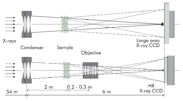
The HRTXM experiment was set up at the micro-optics test bench of beamline ID06 using X-rays from 10 to 20 keV. It makes use of a condenser for sample illumination in imaging mode and as Fourier transformer in diffraction mode. The condenser is comprised of 11 Be parabolic lenses with 300 micrometre radius of parabola apex giving a focal length of ~6 m. The objective lens assembly of 45-62 individual Be parabolic lenses with 50 micrometre radius of parabola apex was located at 2.3 m from the condenser. Two X-ray 2D cameras were used: (i) large area Photonics science CCD detector with 9 µm pixel size for diffraction experiments and (ii) high resolution Sensicam CCD detector with 0.645 µm pixel size. In diffraction mode the direct focussed beam was intercepted in front of the camera by a tungsten beamstop. Switching from the diffraction mode to the imaging mode was achieved by placing the objective lens into the beam, and the chosen camera (Figure 140). The tuneable objective lens provides a magnification between 10 and 25. At maximum magnification a resolution of ~100 nm was achieved. For this benchmark study, a ~70 µm thick single domain grain of Australian gem opal as a prototypical photonic crystal was used.
 |
|
Fig. 140: Conceptual layout of the X-ray microscope. Diffraction (upper panel) and imaging (lower panel) modes. |
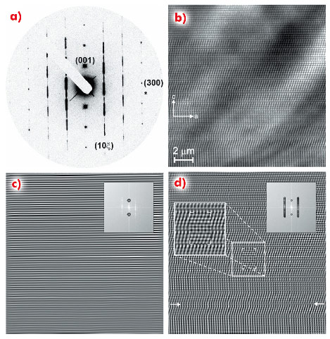
Figure 141 shows a diffraction pattern taken with the X-ray beam parallel to <1-20> zone and the corresponding enlarged X-ray image. Reflections not satisfying the condition H = 3n transform from a well-defined round shape to modulated diffuse rods (see Figure 141a), they are a signature of structural imperfections. The behaviour observed for opal is well known for a close-packed structure with stacking faults, but the surprising feature is the absence of a six-fold axis which must exist for the so-called “growth faults”. The apparent reduction of symmetry of Australian opals to a three-fold symmetry is an indication of unequal probabilities to meet A/B and B/A stackings in the structure. A stripe-like contrast variation with the stripes perpendicular to the c axis is due to a change of the packing sequence (Figure 141b). The orientation of the stripes coincides with striations observable with visible light. Figure 141d shows a filtered HRTXM image, superposed with simulated patterns of a face-centred cubic structure fragment containing a single stacking fault (…ABCABCBCABCABC… sequence) with satisfactory agreement. The most widespread packing mode is obviously f.c.c., but it is not absolutely dominant. A variety of longer-period stacking sequences can be identified, but not being repetitive, they cannot be treated as separate “polytypes” of “phases”.
 |
|
Fig. 141: a) diffraction pattern in <1-20> zone and b) corresponding enlarged high resolution X-ray image. c) and d) Fourier-filtered images, power spectrum with superposed mask sketch is shown as insert. Simulated patterns are given in panel d (see text). Arrows indicate the layer with laterally changing contrast. |
The HRTXM technique, combining high-resolution diffraction and full-field imaging in one setup, provides the tools for efficiently studying the real structure of mesoscopic materials. In structural studies of inverted photonic crystals a resolution in the order of 100 nm was achieved for a wide X-ray energy range from 10 to 50 keV. Short acquisition times with modern area detectors allow the method to be extended to time-resolved studies and combined 3-D real/reciprocal space mapping. The HRTXM method can easily be implemented on existing beamlines. As immediate practical application we mention the characterisation of the real crystal structure during photonic crystal growth.
Principal publication and authors
A. Bosak (a), I. Snigireva (a), K. Napolskii (b) and A. Snigirev (a), Adv. Mater. 22, 3256-3259 (2010).
(a) ESRF
(b) Moscow State University (Russia)
References
[1] A. Snigirev, V. Kohn, I. Snigireva and B. Lengeler, Nature 384, 49-51 (1996).
[2] V. Kohn, I. Snigireva and A. Snigirev, Opt. Comm. 216, 247-260 (2003).
[3] M. Drakopoulos, A. Snigirev, I. Snigireva and J. Schilling, Appl. Phys. Lett. 86, 014102 (2005).
partners


European Synchrotron Radiation Facility - 71, avenue des Martyrs, CS 40220, 38043 Grenoble Cedex 9, France.