- Home
- The impossible fish brain revealed by synchrotron holotomography
The impossible fish brain revealed by synchrotron holotomography
Animal fossils are generally remains of mineralised hard tissues (i.e. bones, teeth or shells), but traces of soft tissues, otherwise destroyed during decay, may be preserved under exceptional circumstances. Accessing this kind of information for extinct organisms becomes increasingly important for our understanding of the evolution of life on our planet. Third generation synchrotrons, and especially the ESRF, now yield evidence of such exceptional soft tissue structures, hitherto regarded as unavailable to palaeontologists. The mineralised brain was discovered in the fossil of a 300 million year-old fish. Findings such as this provide key information about the evolution of life from a very long time ago.
 |
|
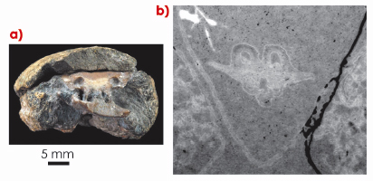
Fig. 142: a) The skull of a 300 million iniopterygian fish from Kansas (USA) preserved in a concretion formed in a reducing shallow water marine environment (foreground). b) bacteria-induced soft tissue preservation of the brain revealed by synchrotron holotomography. |
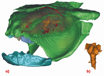
The discovery started with a first test using synchrotron microtomography in absorption mode on the skull of an iniopterygian (an extinct fish group distantly related to present day sharks and ratfish), performed on beamline ID19. Figure 142a shows this specimen which had been rapidly preserved after death in a phosphate concretion. The original purpose of the experiment was to provide the first 3D evidence of the skull for this group of cartilaginous fish which have rarely been preserved in 3D. In addition to the morphology of the skull, the first microtomography test showed a strange, small, and nearly symmetrical object inside the braincase. Despite the insufficient initial resolution and the faint contrast due to the absorption imaging mode, the unexpected structure has similarities with the brain of a modern shark. Exceptional calcification of fossil soft tissues (notably muscles) is already known, but brain and nerve tissues decay so quickly that their preservation was considered impossible. In order to confirm the nature of this object, we decided to apply a far more sensitive method, one that was only recently optimised for such dense specimens: quantitative phase contrast tomography (or holotomography). This method revealed the structure (Figure 142b) in much more detail and with improved contrast. Figure 143a shows the 3D reconstruction of the skull with the enigmatic structure in the middle. The structure displays a morphology characteristic of an early vertebrate brain, with optic lobes, cerebellum, spinal cord and major cranial nerves (Figure 143b). This is the first ever discovery of a mineralised brain replica. The large optic lobes and small cerebellum suggest the habitat of this fish: a bottom-dwelling ambush predator with very large eyes and limited manoeuvrability.
 |
|
Fig. 143: a) 3D segmentation of the same fossil fish skull showing the position of the mineralised brain (orange) and b) dorsal view of the preserved brain. |
How could this exceptional soft tissue preservation occur? Experiments have recently been carried out to understand how fast soft tissues decay in various kinds of organisms, and how bacteria could initiate a rapid transformation of ‘flesh’ into ‘rock’ [1]. ‘Decay experiments’ performed on extant sea urchin embryos demonstrated that, within a few days and under anoxic conditions, bacteria can deposit enough calcium phosphate on the surface of a decaying organic tissue to preserve its shape, until further growth of the crystals makes it resistant to the pressure from the sediment [2].
It appears clearly that the abundance of bacteria, calcium and phosphorus ions, in a very low energy, anoxic, reducing environment, is critical for soft tissue preservation. Therefore, exceptional tissue preservation most frequently occurs near the gut and often concerns muscles of the body wall. The preservation of a brain, although probably due to the same microbial induction, was really unexpected. Our specimen, like many other fossils from the same Carboniferous site in Kansas, may be in fact a regurgitation pellet from a larger shark. The fish may have macerated for a while in the intestine of the shark, where it was enriched with bacteria and phosphorus from other digested prey.
A world specialist of fish brains once declared at a meeting: “I would sell my soul to see the brain of Eusthenopteron” (the 380 million year-old, iconic fossil “intermediate” between fishes and land vertebrates). Our discovery, albeit 80 million years younger, shows that one may not need to go that far to see the “brain of Eusthenopteron”; a synchrotron beamline like ID19 may be enough.
References
[1] D.E.G. Briggs, Ann. Rev. Earth Planet. Sci. 31, 275-301 (2003).
[2] E.C. Raff, J.T.Villinski, F.R.Turner, P.C.J. Donoghue and R.A. Raff., PNAS 103, 5846-5851(2006).
Principal publication and authors
A. Pradel (a,b), M. Langer (c), J.G. Maisey (d), D. Geffard-Kuriyama (a), P. Cloetens (c), P. Janvier (a) and P. Tafforeau (c). PNAS, 106, 5225-5228 (2009).
(a) UMR7207 du CNRS, Muséum National d’Histoire Naturelle, Paris (France)
(b) UPR9034 du CNRS, Gif-sur-Yvette (France)
(c) ESRF
(d) American Museum of Natural History, New York (USA)
partners


European Synchrotron Radiation Facility - 71, avenue des Martyrs, CS 40220, 38043 Grenoble Cedex 9, France.