- Home
- Radiation Therapy using Monochromatic X-rays
Radiation Therapy using Monochromatic X-rays
Review of new developments in fundamental and applied research at ID17 by F. Esteve on behalf of the INSERM U647 RSRM Team Members (INSERM-RSRM CHU/ESRF Grenoble, France)
Despite considerable efforts in cancer therapy, brain tumours like gliomas, are still considered to be some of the most radioresistant tumours. Incidence rate of gliomas is about 5/100 000 and virtually no patient with high-grade glioma survives for more than five years after the disease diagnosis.
The research programs developed by the RSRM team, with the active support of beamline ID17's staff, have explored and confirmed the potential of high-flux monochromatic beams for radiation therapy purposes. At ID17 appropriate infrastructure has allowed the development and the optimisation of innovative chemo-radiotherapy treatments against brain tumours using the particular properties of synchrotron radiation.
Theoretically, to irradiate high-Z elements at their respective K-edge absorption energy leads to Auger electrons and photoelectrons releasing a large amount of energy in their immediate vicinity. For this phenomenon to occur, called photoactivation, requires a powerful monochromatic radiation beam of appropriate energy is required. To date, only synchrotrons allow the production of high-fluence monochromatisable X-rays necessary to reach the above requirements.
We first assessed the enhancement of dose delivered to tumours cells [1], when using the appropriate wavelengths. For instance, when using iodine as a high-Z compound, the optimal energy deposit is obtained close to 50 keV. Extensive cell survival curves studies after irradiation were performed by using various combinations of high-Z compounds, either a simple iodinated contrast agent or chemotherapy drugs containing a heavy element [2] (platinum or iodine). The first preclinical trials were carried out on rats bearing F98 brain glioma using the method previously developed for brain perfusion imaging. These very first preclinical trials [3] have shown that it is possible to master the irradiation procedure with regards to the wavelength choice and to the ballistic (Figure 128), thus leading to an optimal energy deposit in the tumour while sparing surrounding tissues. The same procedure was then used to test the combination of chemotherapy and radiotherapy.
 |
|
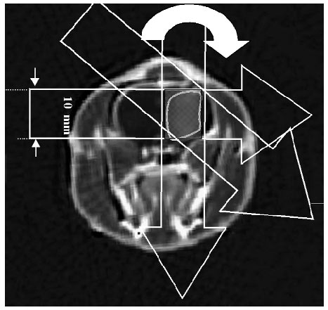
Fig. 128: Irradiation protocol: the tumour is centred on the rotation axis. The beam dimensions are reduced (10 mm wide by 0.8 mm thick). A cylinder of 10.4 mm height and 10 mm diameter is then homogeneously irradiated in tomographic mode. The iodine delineates the tumour volume. When targeted with the appropriate photon wavelength, a dose enhancement effect is caused by the photo electrons emitted by the iodine, leading thus to a better tumour control. |
At the same time, analysis of the onset of DNA damages dependency on the wavelength and the added high-Z compound was also carried out. In vitro experiments were performed with drugs or contrast agents widely used in clinics to check the different achievable radiosensitisations at the DNA scale.
Hence, to irreversibly damage the DNA of tumour cells, an ideal anti-cancer treatment would consist of combining DNA-binding photoactivable high-Z compounds with monochromatic synchrotron irradiation at their K-edge energy. Among the candidate drugs, molecules containing platinum atoms (Z = 78; K-edge energy: 78.4 keV), such as cis-diamminedichloroplatinum (II) (CDDP), are widely used in anti-cancer treatments. The RSRM team has attempted to observe the synchrotron CDDP photoactivation effect in vitro and in vivo, which they have called PAT-Plat. For the first time, a molecular model explaining the biological mechanisms involved in PAT-Plat was proposed and its clinical impact is being investigated [4].
There is now evidence that unrepaired DNA double-strand breaks (DSBs) are the key lesions responsible for cell lethality. In response to radiation, mammalian cells activate two independent DSB repair pathways: 1) the non-homologous end-joining (NHEJ), consisting roughly of the ligation of broken DNA strands. This repair pathway depends upon the protein kinase DNA-PK activity; 2) the homologous recombination, consisting of inserting homologous copies of lacking fragments inside the break. This pathway is mediated by the RAD51 protein, which, when activated, appears in cell nuclei as discrete dots called foci (Figure 129). Unlike radiation, CDDP does not induce DSBs but DNA adducts, generally repaired by a RAD51-dependent recombination. Furthermore, it is known that high concentrations of CDDP inhibit the DNA-PK activity and block the NHEJ repair pathway. Hence, CDDP treatments combined with radiation raise the question of the interplay between two major DNA repair pathways.
 |
|
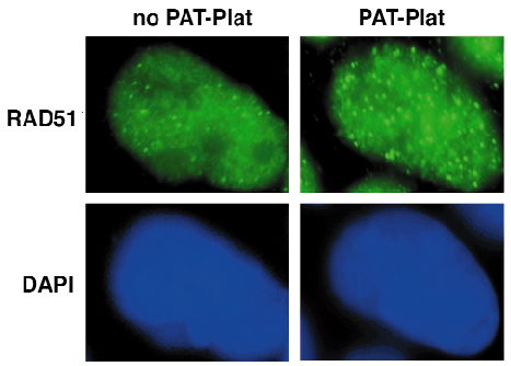
Fig. 129: Wavelength dependency of the DNA damages repair mechanisms. RAD51 nuclear relocalisation in human head and neck carcinoma SQ20B cells treated with CDDP and exposed to synchrotron radiation. Representative examples of RAD51 foci (green dots) in cells irradiated at 78.0 keV (below the platinum K-edge) (left panels) or at 78.8 keV (above the platinum K-edge; PAT-Plat conditions). Cell nuclei are counterstained with DAPI dye (blue). The number of foci is much higher above the platinum K-edge. |
After irradiation at synchrotron X-ray energy above the platinum K-edge (PAT-Plat conditions), CDDP-treated cells showed three times more DSBs than at energies below the platinum K-edge. Furthermore, these DSBs induced in excess at energy above the platinum K-edge were preferentially produced at the close vicinity of the CDDP-induced DNA adducts and were more slowly repaired than in any other conditions [4]. These findings suggest that PAT-Plat produces specifically more complex and more severe DNA damage in CDDP-treated cells than at any other energy. DNA-PK activity was found to be completely deactivated while RAD51 foci appeared more numerous in each cells, as if PAT-Plat effect would result in the inhibition of NHEJ and forces cells to repair by a recombination pathway [4] (see Figure 129). PAT-Plat effect was therefore suggested to be particularly efficient in recombination-deficient cells like tumours. Hence, PAT-plat conditions were naturally applied in vivo to rats bearing glioma. Preliminary studies showed encouraging results with a striking protraction of life span (about 200 days after tumour inoculation) associated with a progressive disappearance of tumour tissue [5]. Our very last experiments led to the most protracted survival (34% at one year) ever reported with this radioresistant glioma model and demonstrate the interest of powerful monochromatic X-ray sources as new tools for cancer treatments [6].
References
[1] F. Estève, S Corde, H. Elleaume, J.F. Adam, A. Joubert, A.M. Charvet, M.C. Biston, J. Balosso, J.F. Le Bas, Academic Radiology, Aug 9 Suppl 2: S540-3 (2002).
[2] S. Corde, PhD thesis, October 2002; and paper submitted to British Journal of Cancer.
[3] J.F. Adam, H. Elleaume, A. Joubert, M.C. Biston, A.M. Charvet, J. Balosso, J.F. Le Bas, F. Estève, Int. J. Radiat. Oncol. Biol. Phys., 57(5), 1413-1426 (2003).
[4] S. Corde, J. Balosso, H. Elleaume, M. Renier, A. Joubert, M.C. Biston, J.F. Adam, A.M. Charvet, T. Brochard, J.F. Le Bas, F. Estève, and N. Foray, Cancer Research 63, 3221-3227 (2003).
[5] M.C. Biston, A. Joubert, H. Elleaume, J.F. Adam, A.M. Charvet, S. Corde, J.F. Le Bas, F. Estève and J. Balosso, 12th International Congress of Radiation Research, Brisbane, (August 2003).
[6] M.C. Biston, A. Joubert, J.F. Adam, H. Elleaume, S. Bohic, A.M. Charvet, F. Estève, N. Foray, J. Balosso, accepted, Cancer Research (2004).
partners


European Synchrotron Radiation Facility - 71, avenue des Martyrs, CS 40220, 38043 Grenoble Cedex 9, France.