- Home
- Local Structure of Spider Silk
Local Structure of Spider Silk
Orb-weaving spiders produce up to 7 different silks, which differ considerably in mechanical properties [1]. We have, however, only very limited knowledge on the structural properties of these silks and how they vary between different species. Scanning X-ray micro-diffraction (SXD) techniques developed at the beamline ID13 can be used to probe single synthetic- and biopolymer-fibres and can thus be applied to large range of silks. We used SXD to study silk produced by the orb-weaving spider Eriophora fuliginea [2]. Orb webs spun by Eriophora are unusually stretchy which contributes to their ability to intercept strong, crepuscular or night flying moths.
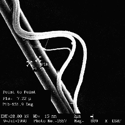 |
Fig. 20: Scanning electron microscopy (SEM) image of Eriophora fuliginea support silk (courtesy: I. Snigireva, ESRF) |
A scanning electron microscopy (SEM)-image of a sample classified as "support thread" shows that it is not a homogeneous material (Figure 20). Two fibres, hypothesised to be of major ampullate (MA) gland origin, are about 7 µm diameter (thick fibres) and form a central thread. A second thread of unspecified glandular origin, made of about 1 µm diameter fibres (thin fibres), is loosely attached to the central thread. It resembles visually, however, silk derived from the minor ampullate glands. An SXD "image" was obtained by mapping the sample through a 3 µm diameter X-ray beam with a 3*4 µm step resolution. After every step, a 2D-diffraction pattern was recorded with a CCD detector. This allows reconstituting an "image" where the "pixels" correspond to individual fibre diffraction patterns with the ß L(polyalanine) structure (Figure 21).
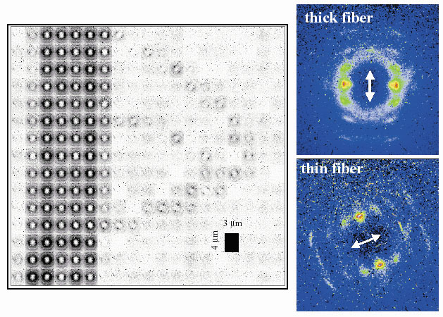 |
| Fig. 21: Scanning X-ray microdiffraction (SXD) image of support silk sample shown in Figure 20. The SXD-image has to be flipped vertically in order to correspond to the SEM image orientation. Individual patterns from the thick and thin fibres are shown on the right. Arrows show the orientation of the macroscopic fibre axis. |
Although the SXD-mapping resolution is less than for a SEM image, one can recognise the essential macroscopic features of the thick and thin fibres. The fibres show a remarkable homogeneity of unit cell parameters, particle size and crystallinity, which suggests that the spider is capable of maintaining control of the spinning process over macroscopic distances. Our data suggest higher volume crystallinity for the thin fibres as compared to the thick fibres. This could be due to a higher amount of crystal-forming polyalanine blocks, as suggested for the fibroins of minor ampullate silk of Araneus diadematus [3]. For the central thread, the orientation of the equatorial reflections is as expected normal to the macroscopic fibre axis. In contrast, the equator is oriented nearly parallel to the macroscopic fibre axis in the thin fibre. To explain this difference, we propose a ribbon-like morphology for the crystalline fraction of the thin fibres. This model implies a tilting of the equatorial line due to the projection of a helical structure on the macroscopic fibre axis. A ribbon-like morphology could also explain the apparent flexibility of the thin fibres.
References
[1] J.M. Gosline, M.E. DeMont and M.W. Denny, Endeavour, 10(1), 37 (1986).
[2] C. Riekel, C.L. Craig, M. Burghammer and M. Müller, Naturwissenschaften, 88, 67 (2001).
[3] P. Guerette, D. Ginzinger, Bhf. Weber and J.M. Gosline, Science, 272, 112 (1996).
Principal Publication and Authors
C. Riekel (a), M. Burghammer (a), C.L. Craig (b) and M. Müller (c), Naturwissenschaften, 88, 67 (2001).
(a) ESRF
(b) Tufts Biotechnology Center, Department of Chemical Engineering, Tufts University, Medford (USA)
(c) Institut für Experimentelle und Angewandte Physik, Kiel (Germany)
partners


European Synchrotron Radiation Facility - 71, avenue des Martyrs, CS 40220, 38043 Grenoble Cedex 9, France.