Introduction
X-ray images are not new: they were probably the main contribution to what we would call today the "mediatic impact" of the discovery of X-rays by Röntgen, just over a century ago. Radiography, or absorption imaging, remains a frequently used and useful technique, either in its original form, or under more elaborate guises such as synchrotron radiation angiography, which uses the subtraction of images around a K-edge.
What is new is the tremendous, and partly unforeseen, development of X-ray imaging techniques that have occurred over the last few years, in connection with the development of modern synchrotron radiation sources. The reasons lie in the association of the source characteristics with the new detectors and computers. They can be described using a few keywords such as "three-dimensional", "high spatial resolution", "coherent beams", "in situ", "real-time", and "combination of techniques". The range of applications is very wide: not only does it includes topics from physics, materials science and engineering, but it also includes those from geophysical, environmental, medical and biological investigations.
The selected articles highlight these different aspects. Furthermore, they show in particular:
- new aspects of Bragg diffraction imaging ("X-ray topography"), with the in situ growth of an epitaxial layer and the real time visualisation of the occurrence of misfit dislocations
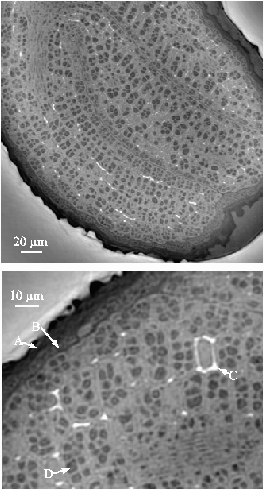
- the importance in microtomography of phase contrast imaging and of quantitative measurements, which are crucial to our understanding of the importance of the mineralisation process in osteoporosis treatments
- the emergence of new ways of scanning imaging, associated with the recent evolutions of microfocussing devices: a new microprobe end-station, ID18F, is devoted to microfluorescence scanning imaging
- the application of X-ray microscopy, using the fluorescence yield, to resolve environment-related problems, such as the intermediate structure within clay gels
- the combination of techniques, which led to new results in X-ray fluorescence microtomography, and to the first combination of Bragg diffraction imaging ("topography") with microtomography, used to reveal the three-dimensional arrangement of dislocations in a diamond crystal.
A growing and important topic concerns all things related to phase contrast images. A few examples are given in the applied and industrial applications section of the present Highlights issue. Let us emphasise here that 1) the use of a coherent beam allowed the combination of Bragg and Fresnel diffraction to access very elusive information about the matching of ferroelectric domains, and 2) that holotomography, which combines phase retrieval (using images recorded at several distances) and microtomography, is now routinely used for a wide variety of problems, which range from the visualisation of the phases in semi-solid alloys to the investigation of fine details of biological materials, (as can be observed in Figure 135) in this case an Arabidopsis plant.
 |
(Courtesy P. Cloetens and R. Mache) |
partners


European Synchrotron Radiation Facility - 71, avenue des Martyrs, CS 40220, 38043 Grenoble Cedex 9, France.