- Home
- Users & Science
- Scientific Documentation
- ESRF Highlights
- ESRF Highlights 2001
- Micro-analysis and Imaging
- Oriented Mesostructures in Aqueous Clay Gels
Oriented Mesostructures in Aqueous Clay Gels
Phase transitions in colloidal suspensions have attracted considerable attention in recent years due to the potential of such systems for fundamental and applied research. The phase behaviour of anisotropic colloids (relevant to most natural systems) is complex due to possible orientational ordering which can lead to inorganic liquid crystals. Suspensions of rod-like particles exhibit an isotropic/nematic phase transition described by Onsager's theory, which also applies to the case of uncharged plate-like colloids as revealed by recent experiments on monodisperse or slightly polydisperse platelets [1]. The situation is much less clear in the case of charged colloidal plates such as natural swelling clays. Indeed clay mineral suspensions do not exhibit a clear isotropic/nematic phase transition with phase separation but, instead, at very low volume fractions (as low as 0.5 wt%) a sol-gel transition turns out to be ubiquitous. However, natural clay minerals are extensively used in drilling fluids because of these gelling properties. The mechanism of gel formation and the resulting gel structure are still not fully understood.
In this regard, direct gel visualisation would represent a significant advance. Multi-keV X-ray microscopy appears as a most promising technique for such a purpose, since it enables the investigation of hydrated samples without any pretreatment. Montmorillonite gels were examined using the X-ray microscopy beamline, ID21. The beam energy was fixed at 2500 eV to ensure a good fluorescence yield for silicon. The investigated depth was estimated to be around 50 µm. A few mg of Wyoming Na-montmorillonite gel (50 g.l-1) were placed in a vacuum-tight cell with two Kapton windows. Figure 141 presents the silicon fluorescence yield image obtained on a montmorillonite gel. The most striking feature of this image is the presence of long range orientational order (~ 200 µm) with aligned domains richer in silicon (3 to 8 µm wide) alternating with Si-poor zones (5 to 30 µm wide) corresponding to water domains. Concentration profiles perpendicular to the lamellae reveal a periodical evolution with a concentration dependent period (Figure 141).
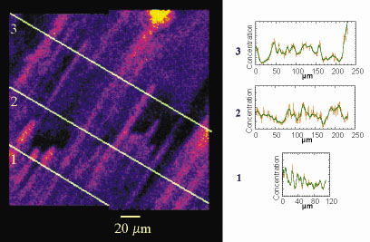 |
Fig. 141: Silicon fluorescent yield mapping of a Na-montmorillonite clay gel at 50 g/l. The image was rebuilt from four different scans 100 mm by 100 µm with a resolution of 1 µm and a dwell time of 400 ms. 1, 2, 3 correspond to three profiles analysed on the right of the figure. |
This image represents the first direct experimental evidence for the existence of a super-structure in montmorillonite gels. It must be stressed that this orientational order is not directly due to individual clay platelets for which the size lies between 0.1 and 0.8 µm, i.e. two orders of magnitude lower than the observed silicon-rich zones, but to "packs" of clay layers, the structure of which remains to be determined. Our preliminary study raises numerous questions about the fundamental physical mechanisms underlying the formation of such entities (isotropic/nematic transition frustrated by charge [2] and/or entanglement, demixtion, spinodal decomposition). In future work, we intend to explore the full phase diagram of concentration and ionic strength by combining X-ray microscopy experiments with optical and rheological measurements. We also would like to study in a more systematic way the influence of charge, particle morphology (size and anisotropy) and polydispersity on the existence and formation of the observed superstructures by combining experiments on synthetic clay samples and on natural montmorillonites from various geological environments.
References
[1] F.M. van der Kooij, K. Kassapidou and H.N.W. Lekkerkerker, Nature, 406, 868 (2000).
[2] A. Mourchid, A. Delville, J. lambard, E. Lécolier and P. Levitz, Langmuir, 11, 1942 (1995)
Principal Publication and Authors
I. Bihannic (a), L.J. Michot (a), B.S. Lartiges (a), D. Vantelon (a), J. Labille (a), F. Thomas (a), J. Susini (b), M. Salomé (b) and B. Fayard (b), Langmuir, 17, 4144 (2001).
(a) Laboratoire Environnement et Minéralurgie, CNRS-INPL-ENSG UMR 7569, Vandoeuvre lès Nancy (France)
(b) ESRF
partners


European Synchrotron Radiation Facility - 71, avenue des Martyrs, CS 40220, 38043 Grenoble Cedex 9, France.