- Home
- Figure 4
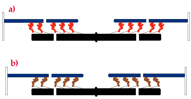
Figure 4
Fig. 4: The two arrays of myosin heads in each half of the myosin filament (black) in the conformation assumed during isometric contraction (a), and in the absence of ATP (b). Actin filaments shown in blue.
| back to: X-ray Interference Reveals Angstrom-Scale Motions of Myosin in Intact Muscle Cells |
partners


European Synchrotron Radiation Facility - 71, avenue des Martyrs, CS 40220, 38043 Grenoble Cedex 9, France.