- Home
- Figure 3
Figure 3
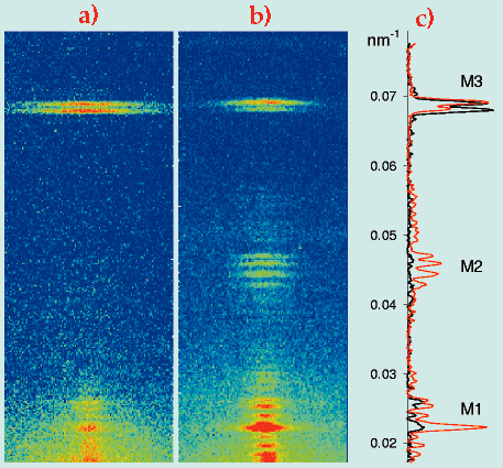
Fig. 3: Axial X-ray diffraction pattern recorded at ID2 from a single fibre isolated from frog muscle, during active contraction (a, 6s exposure) and in the absence of ATP (b, 60s exposure) and comparison of the respective axial intensity distributions (c; active, black; rigor, red).
| back to: X-ray Interference Reveals Angstrom-Scale Motions of Myosin in Intact Muscle Cells |
partners


European Synchrotron Radiation Facility - 71, avenue des Martyrs, CS 40220, 38043 Grenoble Cedex 9, France.