Sample preparation
BM29 Sample Information
Information about the lab equipment and procedures (including protein standard preparation and concentration measurement) useful for the sample preparation, the day of the BioSAXS experiment.
Sample preparation lab
The sample preparation laboratory is in 30.0.06 room located on the outside side of Experimental hall in front of ID30 MASSIF. 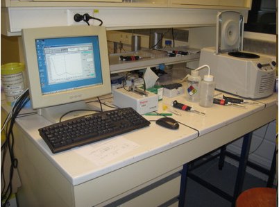
In the sample preparation room, you will find:
- desktop Centrifuge thermostated from 0 deg. C to 40 deg. C (an ultracentrifuge with a unique rotor (TLA55), 12 places and a maximum volume of 1.5 mL is available at the EMBL laboratory upon request in the A-form, a cooled centrifuge for 15 and 50 mL tubes is temporally located in Experimental hutch)
- Nanodrop 1000, Nanodrop ONE and Epoch Plate Reader (possible reading at several wavelengths)
- Fridge (4 deg.C) with freezer drawers at -20 deg.C and a freezer at -80 deg. C
- Ice machine (dry ice available on the site upon request)
- Magnetic steerer
- Balance, a MiliQ, pipettes, tips, plates, eppendorfs and PCR tubes, etc.
Preparing a BSA sample
Each user group prepares at beginning of its beamtime a BSA sample. The aim is to learn/be confident how to use sample prep facilities, sample changer and carry out an experiment (beamline hardware and software).
The buffer for the BSA (50 mM HEPES pH 7.5) and the BSA powder are in the bottom drawer of the fridge in the sample preparation lab.
-
- Weigh 5 mg of BSA in an eppendorf tube and add 1 ml of buffer.
- Mix by pipetting (try to do it calmly to minimize foam formation).
- When it is completely dissolved centrifuge it in the cooled room centrifuge at 4°C, 14000 rpm for 15 minutes.
- Put the supernatant in a fresh tube (it is quite possible that no pellet can be seen, but, to be safe, avoid touching with the tip the bottom of the tube while transferring to the new eppendorf).
- Measure the concentration of the solution at the Nanodrop spectrophotometer (instructions below). Usually, you should expect concentrations between 4 and 5 mg/mL.
Water as Absolute Reference
Local contact of the day measure water (empty capillary + water) for users as an abolute reference and modify normalisation coefficient (a number which normalise each pixel value) if necessary. The molecular mass of samples (proteins, nucleic acids, protein-nucleic acid complexes, lipids, carbohydrates,etc...) can be than estimated using the known absolute scattering of water at a given temperature and athmospheric preasure.
I0,abs (water)= 1.632.10-2 cm-1, at 20 deg. C
Please, find here, an Excel file .xls which allows for calculating the MM of your samples (monomers or complexes) using water as reference: Molecular Weight Calculation
The experimental parameters to modify into the xls file are only:
- the partial specific volume of your sample (cm3/g)
- the zero scattering intensity of water (relative unit) given by your local contact the day of the experiment
- the zero scattering intensity of your samples (in relative unit) obtained after the BioSAXS measurements.
The intensity can be also experesed in absolute scale cm-1 upon request.
Measurement of the sample concentration
In practice, inaccuracies on concentration measurements are larger sources of errors in molecular mass estimation than the precision of calculating I0 itself [Feigin & Svergun (1987)].
For the molecular mass estimation, the knowledge of the sample concentration is crucial. We use either a Nanodrop or a plate Reader to measure the concentration of our samples (2 µL of sample are enough). We recommend measuring the concentration right after sample centrifugation.
Using the Nanodrop spectrophotometer
- Open the software by clicking computer double icon nd-1000
- To measure the protein concentration at 280 nm press the “Protein A280” button.
-
In “Sample type” box, select “1 Abs = 1mg/ml” or “Other protein (E & MW)” and “Other protein (E 1%)” if you are measuring the concentration of a protein knowing its extinction coefficient).
-
Press “Blank” after placing 2 µl of buffer
-
Press “Measure” after putting 2 µl of the protein sample (usually concentrations up to 50 mg/ml can be handled without dilution).
The Nanodrop measures then the absorbance (optic density, OD) at 280 nm and converts it into a concentration using the Beer-Lambert relation (pathlength=10 mm).
partners


European Synchrotron Radiation Facility - 71, avenue des Martyrs, CS 40220, 38043 Grenoble Cedex 9, France.