- Home
- Users & Science
- Scientific Documentation
- ESRF Highlights
- ESRF Highlights 2013
- Structure of materials
- Watching ice crystals growing in colloidal suspensions
Watching ice crystals growing in colloidal suspensions
Ice crystals growing in a colloidal suspension is a phenomenon encountered in a variety of natural processes such as the freezing of soils in northern regions and the growth of sea ice, or everyday life and engineering situations such as frozen dessert processing, materials science, cryobiology, filtration or water purification, and the removal of pollutants from waste.
The phenomenon by itself is surprisingly simple to describe: a solidification interface, usually the water/ice interface, is propagating through a colloidal suspension of particles, cells or micro-organisms. This simplicity is nevertheless misleading and we are still far from a correct understanding and control of the phenomenon. If the various manifestations of colloid freezing can be observed in numerous natural or technological occurrences, precise and quantitative observations are required in order to understand the process. Current approaches, both direct such as optical microscopy and X-ray radiography and indirect such as X-ray scattering only provide partial, averaged or indirect observations.
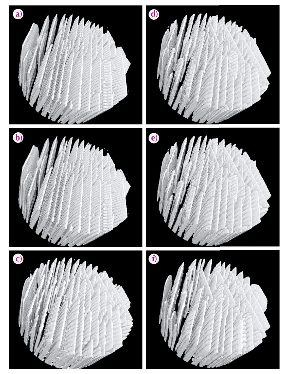
The velocity range of interest (1–50 µm.s–1), corresponding to the usual occurrences of colloids freezing, nevertheless requires high resolution, fast imaging capabilities. Here, we demonstrate that fast X-ray computed tomography allows time-lapse, three-dimensional in situ imaging of ice crystal growth in a colloidal silica suspension. We took advantage of recent upgrades and developments at beamline ID15, which, due to the high flux of high energy X-rays available, allows us to perform a complete tomographic acquisition at high resolution (voxel size: 1.786 x 1.786 x 1.786 µm3) within a second, providing thus a time-lapse view at ice crystals growing in a colloidal suspension (Figure 122).
 |
|
Fig. 122: Time-lapse sequence of ice crystals morphology. 3D reconstruction of the ice crystals, from tomographs taken every minute. The diameter of the reconstruction region is 860 µm. The colloids, concentrated between the growing crystals, are not shown on the reconstruction. Voxel size: 1.786 x 1.786 x 1.786 µm3. |
Three dimensional in situ observations of such solidification systems have never been obtained so far. What is new and unique here can be placed in three main categories:
- Qualitative observations: the actual morphology of the ice crystals growing into a colloidal suspension have never been observed before. Our observations reveal, for instance, the specific morphology of the tip of the crystal, which is strongly asymmetric (inset in Figure 123). This is very different from the usual dendritic morphologies observed in solidification, where the tip is highly symmetrical.
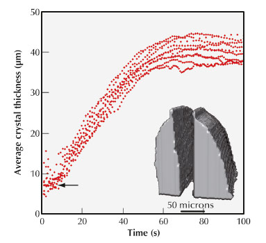
- Quantitative observations: the growth kinetics can be measured both along the temperature gradient and perpendicular to the temperature gradient, a measurement that has never been obtained thus far. The measurements (Figure 123) show that for the latter, the growth kinetics is constant until the particle concentration in the inter-crystals space becomes really high. The presence of particles in this case has thus very little influence over the growth kinetics, until the particles are closely packed. We can also compare the growth kinetics in both directions (parallel and perpendicular to the temperature gradient) and conclude that they are actually quite similar. This is an important result since the lateral growth kinetics dictates the conditions for particle redistribution between the crystals, which in turn influence the behaviour or properties of the frozen assembly.
- X-ray computed tomography is a very popular and powerful technique for solidification studies. These results also highlight some limitations of the technique for this very particular and sensitive system, in particular regarding the influence and absorption of the beam. We found here that aqueous systems are highly sensitive to small variations of temperature (a few degrees), which can drastically affect the solidification behaviour and resulting morphologies. Further improvement of the imaging conditions should nevertheless allow us to obtain artefact-free observations.
 |
|
Fig. 123: Evolution of ice crystal thickness. The corresponding crystals tip morphology is shown in the inset. |
There are currently no alternatives to this fast-tomography imaging approach permitting such qualitative and quantitative information to be obtained. This approach proved critical for our understanding of the complex process of crystallisation from a colloidal suspension.
Principal publication and authors
S. Deville (a), J. Adrien (b), E. Maire (b), M. Scheel (c) and M. Di Michiel (c), Acta Materialia 61, 2077–2086 (2013).
(a) Laboratoire de Synthèse et Fonctionnalisation des Céramiques, UMR3080 CNRS/Saint-Gobain, Cavaillon (France)
(b) Université de Lyon, INSA-Lyon, MATEIS CNRS UMR5510, Villeurbanne (France)
(c) ESRF
partners


European Synchrotron Radiation Facility - 71, avenue des Martyrs, CS 40220, 38043 Grenoble Cedex 9, France.