- Home
- Users & Science
- Scientific Documentation
- ESRF Highlights
- ESRF Highlights 2009
- Enabling Technologies
- MAXIPIX, a fast readout photon-counting pixel detector
MAXIPIX, a fast readout photon-counting pixel detector
X-ray scattering techniques such as XPCS, surface scattering, and SAXS require a low noise 2D detector capable of covering a large range of time and intensity scales. To meet these requirements, we have developed “MAXIPIX”, a fast readout photon-counting pixel detector based on the Medipix2 [1] photon-counting readout chip developed by CERN.
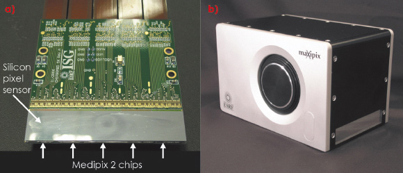
Besides the photon-counting detection mode that provides essentially noise-free data, Medipix2 has other desirable qualities such as a readout dead time of less than 300 microseconds and a small pixel pitch of 55 x 55 micrometres square. These qualities make it particularly suitable for applications requiring high temporal and/or spatial resolution. To make best use of these capabilities, we developed a specific readout board called “PRIAM” for the simultaneous readout of five Medipix2 chips at their maximum speed ratings. We also developed a range of detector modules with flex cable connection to the high bandwidth PRIAM board input ports. Each module implements one or several Medipix2 chips bump-bonded to a 500 µm thick monolithic silicon pixelated sensor. Available detection surfaces are 14 x 14 mm2 (single chip), 28 x 28 mm2 (2x2 chips), and 71 x 14 mm2 (5x1 chips) (Figure 154a). The resulting frame rate is more than 1 kHz with a single chip and up to 350 Hz with multichip modules. The PRIAM board, the detector module and their power supplies are hosted in a compact housing, resulting in the MAXIPIX detector (Figure 154b). MAXIPIX is operated using a Linux-based high data rate acquisition station developed by the ESRF and achieving 1.6 Gbit/s data transfer rates.
 |
|
Fig. 154: a) Multi-element detector module with 5 Medipix2 chips and 1280 x 256 pixels. b) MAXIPIX detector head. |
The short readout dead time is exploited not only for high frame rate acquisition, but also for acquisition of high statistics data by frame accumulation with virtually no dead time losses.
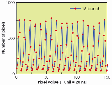
The PRIAM readout board is also compatible with Timepix [2], a version of Medipix2 implementing a time stamping mode in addition to the standard photon-counting mode. This allowed clear resolution of the 176 ns period of the 16-bunch mode of the ESRF source (Figure 155) and opens new possibilities for time-resolved 2D detection in the 100 ns time range.
 |
|
Fig. 155: Histogram of pixel values in images taken with MAXIPIX/Timepix in16-bunch machine mode, showing the time structure of the beam. |
The MAXIPIX development was focused on high time and space resolutions rather than on a large physical detection area, thereby complementing other photon-counting systems like XPAD or PILATUS.
MAXIPIX is already in use at several ESRF beamlines. The principal applications are inelastic scattering (ID16), XPCS (ID10A), surface diffraction (ID03) and scattering (BM5, ID10B), coherent nanodiffraction (ID01), and micro-SAXS (ID13). In particular, the frame rate and spatial resolution characteristics proved to be particularly well suited to dynamic XPCS experiments [3], whereas the small pixel size together with the noiseless detection was exploited to improve the analyser performance on ID16 [4].
One limitation is the insufficient X-ray absorption efficiency of the silicon sensor above 20 keV. Hence our main development activity at present is focused on pixel sensors based on high-Z semiconductor materials in order to raise the energy limit to 50 keV or more. Recent tests on Medipix2 prototype modules with CdTe and GaAs sensors have shown the rapid advances made in high-Z sensor technologies. We expect to be able to deploy high-Z MAXIPIX versions to beamlines shortly. This will be of benefit to various existing applications. Moreover, it will open new perspectives, in particular in materials science and medical imaging.
In summary, we have built a versatile detector which is now routinely used on beamlines and which enabled improvements in several experimental methodologies. This provides motivation for further development programmes intended to widen the range of its applications.
Acknowledgement
The authors wish to thank the Medipix2 collaboration and in particular X. Llopart and L. Tlustos (CERN) for their advice on Medipix2 chip operation and their support in Medipix2 multichip assemblies testing.
References
[1] X. Llopart, M. Campbell, R. Dinapoli, D. SanSegundo and E. Pernigotti, IEEE Trans. Nucl. Sci. 49, 2279-2283 (2002).
[2] X. Llopart, R. Ballabriga, M. Campbell, L. Tlustos and W. Wong, Nucl. Instr. and Meth. A 581, 485 (2007) and erratum Nucl. Instr. and Meth. A 585, 106 (2008).
[3] C. Caronna, Y. Chushkin, A. Madsen and A. Cupane, Phys. Rev. Lett. 100, 055702 (2008).
[4] S. Huotari, G. Vankó, F. Albergamo, C. Ponchut, H. Graafsma, C. Henriquet, R. Verbeni and G. Monaco, J. Synch. Rad. 12, 467-472 (2005).
Principal publication and authors
C. Ponchut (a), J. Clément (a), J.M. Rigal (a) E. Papillon (a), J. Vallerga (b), D. LaMarra (c) and B Mikulec (d), Nucl. Instr. and Meth. A 576, 109 (2007).
(a) ESRF
(b) University of California Berkeley (USA)
(c) University of Geneva (Switzerland)
(d) CERN (Switzerland, France)
partners


European Synchrotron Radiation Facility - 71, avenue des Martyrs, CS 40220, 38043 Grenoble Cedex 9, France.