- Home
- Users & Science
- Scientific Documentation
- ESRF Highlights
- ESRF Highlights 2008
- X-ray imaging and optics
X-ray imaging and optics
Introduction
The importance of X-ray imaging techniques for innovative research is now widely recognised among the synchrotron radiation user communities. A number of X-ray imaging beamlines are being built in new synchrotron facilities that are emerging all around the world. Two new biomedical beamlines have entered into operation at the end of 2008, in Canada and Australia. The upgrade programmes of major facilities such as the ESRF and APS strongly emphasise X-ray imaging as one of the five “highlight areas” and one of the two “compelling avenues for research” respectively.
This gain in visibility results from the increased number of scientific communities that use X-ray imaging techniques for their research, as reflected by the present chapter. The highlight contributions originate from several scientific areas, which include:
• Biomedical applications, with the investigation of novel contrast agents, an improved knowledge of the effect of microbeams, potentially very important for clinical radiotherapy applications, and the characterisation of stem cells using infrared spectromicroscopy.
• Materials Science studies, with the observation of intergranular stress corrosion cracking in a grain-mapped polycrystal, and the local structure of GaGdN, investigated with a microprobe beam and several complementary techniques.
• Cultural Heritage, where synchrotron techniques revealed the unexpected presence of oil in very ancient paintings of the Bamiyan Buddhist murals, the presence of relics hidden in Middle Age artistic objects, and where phase contrast imaging was used for paleontological purposes, to study fossil evidence hidden in Early Cretaceous opaque amber.
• Environmental Sciences with the investigation, using a 80 nm x 90 nm spot, of cometary grains collected during the NASA Stardust mission, or helping to characterise the impact of carbon nanotubes on human health.
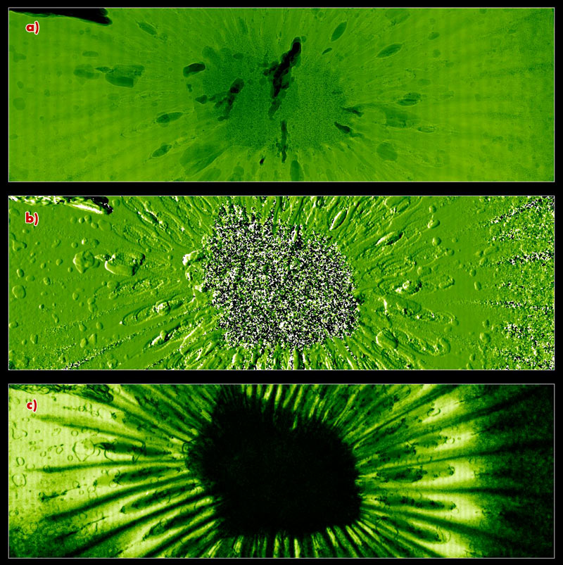
Another point we wish to emphasise is the importance of the effort, made by the ESRF staff and users, to implement new (or new combinations of) imaging techniques, opening new ways for scientific research. This is exemplified, by the first results obtained with an improved diffraction tomography method, as well as the biomedical phase contrast images provided by a grating inteferometry-based technique. The latter can be applied to a wide variety of materials: Figure 125 shows a slice of kiwi fruit in different contrast modes, obtained from a single phase-stepping interferogram series.
 |
|
Fig. 125: Grating interferometric radiographs of a slice of kiwi fruit (ID19, 27 keV): a) absorption image; b) differential phase contrast image (the scattering is so strong in the centre that the phase is not well defined); c) dark-field (i.e., scattering-contrast) image, showing features that are not visible in the absorption and the phase images. The field of view of each frame is 36.5 mm wide and 11.5 mm high; the detector pixel size was 30 µm (Image courtesy: T. Weitkamp, E. Reznikova, C. David et al.). |
The trends, challenges and new opportunities associated with synchrotron radiation-based imaging are clearly defined for the next few years; if we wish to be able to contribute substantially to emerging topics in areas like nanosciences, health and environment preservation, or cultural heritage, we must improve both the spatial (towards the nanometre scale) and temporal resolutions (for the investigation of evolving systems), and be able to achieve higher chemical sensitivity for micro- or nano-analysis. In addition, we have to pursue the implementation of new imaging techniques and the in situ combination of techniques. All this will be performed in a highly competitive international context.
The ESRF Upgrade Programme, where X-ray imaging plays a substantial role, intends to give an answer to these trends and challenges, through several projects, which include very long (up to 200 m) beamlines for Nano Imaging and Nano Analysis (NINA), and parallel and coherent beam Micro-Imaging Applications (MIA). In addition, the next two years will be crucial for the implementation of clinical radiotherapy programmes, at the recently refurbished biomedical beamline. All these upgrades, coupled with the scientific know-how already acquired using X-ray imaging at the ESRF, will constitute a great incentive for both the ESRF staff and users to push the present limits of the techniques to reach new, currently unobtainable, scientific heights.
J. Baruchel
partners


European Synchrotron Radiation Facility - 71, avenue des Martyrs, CS 40220, 38043 Grenoble Cedex 9, France.