- Home
- News
- General News
- X-rays reveal important...
X-rays reveal important mechanism behind parkinsonian disorders
16-01-2019
An inherited gene defect results in manganese poisoning inside cells, which leads to parkinsonism. This is the result of a new study by scientists from CNRS, University of Texas in Austin, DESY and the ESRF.
Parkinsonism is a syndrome that includes a group of disorders affecting the nervous system, which lead, among other symptoms, to a characteristic tremor known as shaking palsy. Globally, 1% of those over the age of 60 are affected by Parkinson's disease. In most cases, it is not possible to determine what brings on the disorder. Apart from Parkinson's disease, there are a number of symptomatic parkinsonian syndromes that can be triggered by very different causes. For instance, manganese, although essential in small concentrations, is thought to be a possible cause of certain types of neurological disorders in higher concentrations.
A team of scientists from the CNRS and University of Bordeaux, the University of Texas in Austin, DESY and the ESRF has revealed one of the key mechanisms behind certain familial parkinsonian disorders. Using synchrotron X-rays they have shown in detail how poisoning with metal manganese occurs inside a cell, leading to parkinsonian symptoms. It found that a specific genetic defect results in manganese accumulating in the cell’s so-called Golgi apparatus. “An exact understanding of this manganese poisoning is a crucial step towards designing possible therapies,” explains the main author of the study, Richard Ortega, scientist at the CNRS.
 |
|
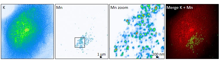
The picture shows the manganese distribution in a mutant cell. Credits: R. Ortega. |
The scientists focused on a mutation in the SLC30A10 gene, previously identified in a series of cases of familial Parkinsonism and which appears to lead to genetically inherited disorders. “SLC30A10 is responsible for transporting manganese out of the cell,” explains Somshuvra Mukhopadhyay from the University of Texas in Austin, one of the co-authors, who identified this function of the gene and the corresponding protein in an earlier study. “In the mutated form, this transport is disrupted so that toxic concentrations of the metal accumulate in the cell.”
The scientists wanted to locate the manganese inside the cell in order to do this; they carried out micro X-ray fluorescence imaging at room temperature at DESY on mutated and non-mutated samples. They found that manganese lit up particularly in the Golgi apparatus of the mutated cells.
The Golgi apparatus is an organelle within the cell that acts as a dispatch centre for proteins: it receives the protein molecules from the endoplasmic reticulum, modifies some of them and then attaches the equivalent of an address label for their further dispatch. The proteins are then carried off in tiny sacs known as vesicles.
With the aim to see where exactly the manganese was, they used cutting-edge experiments of nano X-ray fluorescence imaging of frozen-hydrated samples at the ESRF’s ID16A beamline. “It is the only place where we could spot such small amounts of manganese in nanometric vesicles, thanks to the impressive sensitivity and spatial resolution of ID16A beamline“, says Asuncion Carmona, scientist at CNRS and first author of the article. “Carrying out the high resolution experiments on frozen cells at very low temperature is mandatory, but very challenging. Everything went smoothly thanks to the quality of the new ESRF instrumentation and the expertise of the whole team”, adds Peter Cloetens, in charge of ID16A.
The experiments showed that the manganese collects principally inside the transport vesicles of the Golgi apparatus. The scientists suspect that this disrupts protein transport out of the cell, in particular, which in turn impairs nerve function and leads to the observed parkinsonian symptoms. A corresponding accumulation of the metal was not observed in the Golgi apparatus of healthy cells.
This research focuses on only one of several known mutations of the gene in question and the cells used come from the outer tissue of a cancer growth. “Our goal for the future is to study this manganese accumulation in more detail and in conditions that resemble more closely those under which the disease occurs, for example in suitably mutated nerve cells”, concludes Ortega.
Reference:
Carmona, A. , et al; ACS Chemical Neuroscience, 2018; DOI: 10.1021/acschemneuro.8b00451
Top image: SXRF microprobe imaging of manganese (Mn) in the cells. Credit: R. Ortega.
partners


European Synchrotron Radiation Facility - 71, avenue des Martyrs, CS 40220, 38043 Grenoble Cedex 9, France.