- Home
- News
- General News
- Unveiling the structure...
Unveiling the structure of ‘living electronics’ produced by bacteria
28-09-2026
Researchers have uncovered the molecular architecture of cable bacteria, which transport electricity over centimetre-scale distances. Nano-X-ray fluorescence imaging at the ESRF’s ID16A beamline showed that the bacteria’s conductive fibres contain continuous nickel-rich nanoribbons and provided the quantitative data that pinned down their structure: a naturally occurring metal-organic framework, unlike anything previously known in biology. The results are out in Nature Communications.
Share
Cable bacteria are living electrical cables made of thousands of microbial cells joined end to end. Found in the sediments of rivers, lakes and coastal seabeds, these organisms transport electrical currents over distances of several centimetres, an enormous distance for a bacterium. The current runs through 20 to 70 protein fibres, each about 50 nanometres wide, that run in parallel along the entire filament. Yet since their discovery some fifteen years ago, the molecular basis of this conductivity has remained a mystery.
Now, an international team led by the University of Antwerp has identified the structures responsible for this long-range electron transport. Their study reveals that the conductive fibres inside cable bacteria contain bundles of nanoribbons arranged into a metal-organic framework, a class of material in which metal ions act as nodes and organic molecules as linkers. Metal-organic frameworks, or MOFs, were the subject of the 2025 Nobel Prize in Chemistry and are studied intensively for gas storage, catalysis and electronics. They had never before been found in a living organism.
 |
|
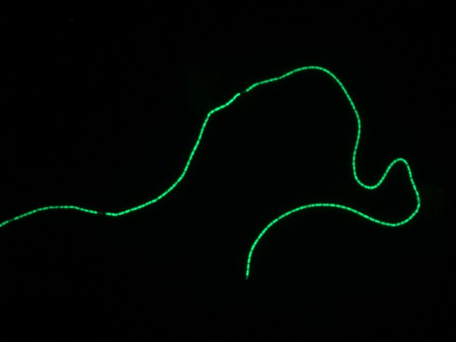
Cable bacteria are multicellular bacteria forming centimeter-long filaments that consist of up to > 10.000 cells in a row. Here is a single filament with a DNA stain. Image credit: Silvia Hidalgo-Martinez (University of Antwerp) |
“We were totally stupefied when we found out that a MOF structure was present in our bacteria”, explains Filip Meysman, professor at the University of Antwerp and lead author of the study. “Somehow evolution came up with the exact same idea as the Nobel Prize chemists.”
Using a combination of nano-X-ray fluorescence imaging at the ESRF’s ID16A beamline, electron microscopy, Raman spectroscopy, electron paramagnetic resonance, X-ray absorption spectroscopy and computational modelling, the researchers showed that the conductive fibres contain intertwined nanoribbons enriched in nickel and sulphur.
Their analyses indicate that these structures are composed of repeating nickel bis(dithiolene) units, referred to as NiBiD complexes. The nickel centres are linked by sulphur-containing ligands to form elongated, highly conjugated molecular chains. These chains align and stack together into nanoribbons that run along the length of the fibres, creating one-dimensional pathways for charge transport.
Mapping the nickel in a living wire
At ID16A, the ESRF’s nano-imaging beamline, the researchers scanned an X-ray beam focused to 35 nanometres across individual bacterial filaments and across “fibre skeletons”, filaments from which the membranes and cell contents had been removed so that only the fibre network remained. The samples were kept frozen at 100 K throughout to protect them from radiation damage. The resulting element maps show nickel concentrated in evenly spaced parallel lines that match the fibres one by one and run unbroken across several cell-to-cell junctions. In the fibre skeletons, sulphur follows the same pattern. The junctions themselves hold a second, copper-rich complex, located in the wheel-shaped structure that electrically connects the fibres of neighbouring cells.
Because X-ray fluorescence measures the absolute amount of each element, the maps did more than locate the nickel. They yielded the nickel content per length of fibre and the sulphur-to-nickel ratio. The first number tells how many nickel chains are packed into each fibre and each nanoribbon; the second rules out one of the two candidate molecular structures suggested by spectroscopy. The molecular model of the conductive fibre is built on these ESRF measurements.
Dmitry Karpov, assistant professor at Université Grenoble Alpes (CEA-IRIG) and former postdoctoral scientist at the ESRF’s ID16A beamline, explains: “The fibres of cable bacteria are so small that we had to push the beamline to its limits in both resolution and sensitivity. Several things had to come together: the extra flux from the EBS upgrade, the cryogenic sample environment of ID16A, which lets the sample survive all those photons packed into a 35-nanometre beam while we scan slowly enough to collect the signal we need, and the long-term stability of the beamline that makes such scans possible at all. And it all had to be quantitative.”
High-resolution electron microscopy at the University of Antwerp exposed the nanoribbons themselves: about eleven per fibre, 1.4 nanometres thick, weaving along the centre of each fibre. X-ray absorption spectroscopy at Diamond Light Source (UK) confirmed that the nickel atoms occupy a square-planar coordination environment characteristic of nickel bis(dithiolene) compounds, closely resembling synthetic conductive coordination polymers studied for organic electronics.
The resulting architecture resembles the braided copper wire in a household power cord: many thin conductors twisted together inside a protective sheath, yet here a layer of protein. Cable bacteria are motile and bend sharply, and an entangled bundle stays conductive while remaining flexible.
The conductivity of these biological structures is exceptional. Earlier measurements put the conductivity of a whole fibre at 5 to 500 S/cm, rivalling the conductive inks used in printed electronics. When transport is considered within the nanoribbon network alone, which fills only a small fraction of the fibre cross-section, the estimated conductivity reaches 150 to 15,000 S/cm, ranking among the highest values reported for any metal-organic material.
Sustainable electronic materials
Beyond solving a long-standing biological puzzle, the findings suggest a new strategy for designing sustainable electronic materials. Nature appears to have evolved efficient metal-organic architecture capable of guiding electrical currents through lightweight, carbon-based structures with only modest amounts of metal.
“Effectively, the cable bacteria turn out to be excellent chemists”, adds Meysman. “Somehow evolution has brought forward a very flexible and highly performant MOF material. This not only opens possibilities for the bio-based synthesis of metal-organic frameworks, but also suggests a novel design principle for sustainable electronic materials.”
Reference:
Meysman, F.J.R. et al. Highly efficient long-range conduction through a biosynthetic nickel-organic framework. Nature Communications 17, 8814 (2026). https://doi.org/10.1038/s41467-026-76989-0
Text by Montserrat Capellas Espuny
Top image: Cable bacteria are multicellular bacteria forming centimeter-long filaments that consist of up to > 10.000 cells in a row. The image shows multiple filaments recorded by polarized microscopy. Image credit: Eric Boschker (University of Antwerp)
partners


European Synchrotron Radiation Facility - 71, avenue des Martyrs, CS 40220, 38043 Grenoble Cedex 9, France.