- Home
- Education & Outreach
- What is the ESRF?
- What is synchrotron light?
The ESRF produces synchrotron light with wavelengths ranging from gamma rays to infrared radiation. It consists mostly of X-rays with a wavelength of about 0.1 nanometre (a nanometre is one billionth of a metre, i.e. 1 nm = 10-9 m).

What are X-rays and why use them?
X-rays were discovered by Wilhelm Röntgen in 1895.
They are electromagnetic waves like visible light but situated at the high energy/short wavelength end of the electromagnetic spectrum, between ultraviolet light and gamma rays. Their wavelength ranges from 0.01 nm to 10 nm, which is comparable to interatomic distances.
Today, X-rays are used extensively in medical imaging because they have a high penetration depth through materials and are selectively absorbed by the parts of the body with the highest electron density such as bones. However, this interesting property is not the only reason why we use X-rays at the ESRF.
In visible light and with the help of an optical microscope, it is possible to observe objects the size of a microbe. However, to be able to "see" atoms, which are 10 000 times smaller, we need light with a very short wavelength. In other words, we need X-rays.
Brilliance and other properties
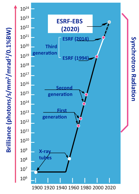
The main difference between synchrotron light and the X-rays used in hospitals is the brilliance: a synchrotron source is one hundred billion times brighter than a hospital X-ray source. The higher the brilliance, the more precise the information that can be obtained from the X-ray.


The synchrotron X-ray beam can have other valuable properties, including time structure (so that it flashes), coherence (making it a parallel beam) and polarization.


